
|
Fig. 3
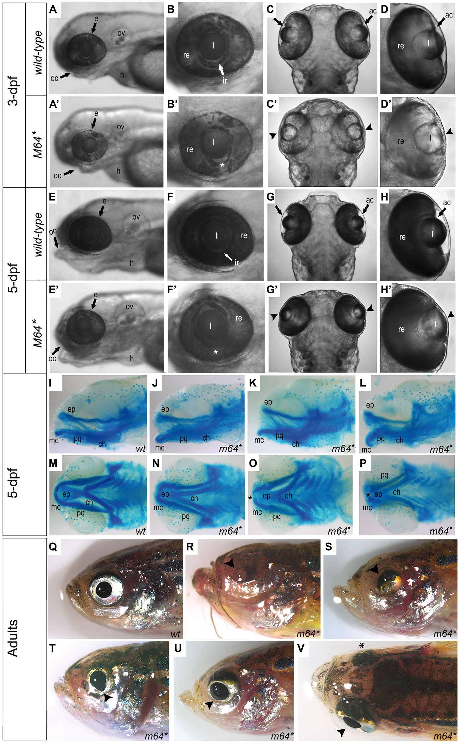
Gross characterization of the pitx2M64* mutant phenotype. (A–H’) Wild-type (A–H) and pitx2M64* mutant (A’–H’) embryos. At 3-dpf, prominent features include ventral iris coloboma (white asterisk in B’ versus B) and bilateral severely underdeveloped ocular anterior chambers (arrowheads in C’, D’ versus C, D). By 5-dpf, in addition to the persistence of the coloboma (white asterisks in F’ versus F) and underdeveloped anterior chambers (arrowheads in G’, H’ versus G, H), mutant lenses of homozygous pitx2M64* embryos appear elongated in the anterior/posterior plane (G’, H’ versus G–H). Craniofacial abnormalities also become apparent in mutants (E’ versus E). Views are lateral (A, B, A’, B’, E, F, E’, F’) and dorsal (C, D, C’, D’, G, H, G’, H’). (I–P) Alcian blue staining of 5-dpf wild-type (I, M) and pitx2M64* mutant (J–L, N–P) embryos. A range of mild to severe structural malformations were evident in the Meckel’s cartilage, mc, palatoquadrate, pq, (first pharyngeal arch) and ceratohyal, ch, (second pharyngeal arch) (I versus J–L, M versus N–P). Views are lateral (I–L) and ventral (M-P). (Q–V) Wild-type (Q) and pitx2M64* mutant (R–V) adult fish. Gross ocular phenotypic presentation included anophthalmia (R), microphthalmia (S), and enlarged/misshapen pupils with improper globe orientation (T, U) and could appear asymmetrically (as in V; arrowhead versus asterisk). Views are lateral (Q–U) and dorsal (V). ac, anterior chamber; e, eye; h, heart; ir, iris; l, lens; oc, oral cavity; ov, otic vesicle; re, retina; ep, ethmoid plate; mc, Meckel’s cartilage (P1); pq, palatoquadrate (P1); ch, ceratohyal (P2); hs, hyosympletic (P2).