
|
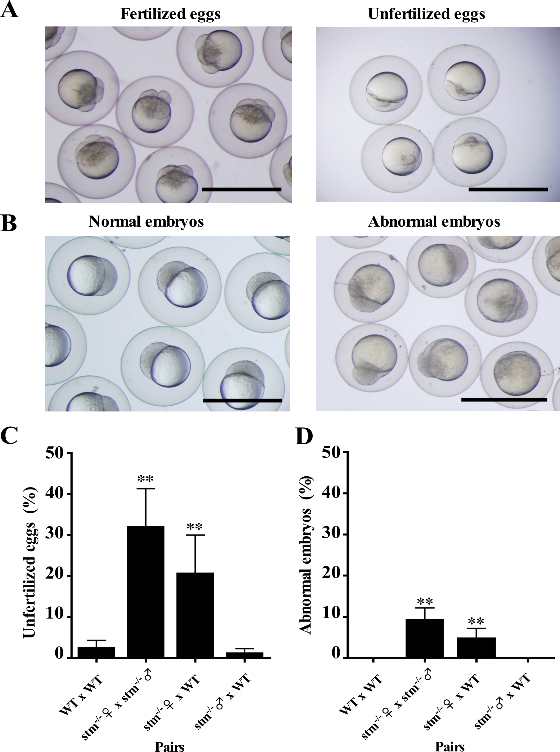
Fig. 6
Morphology and rate of development of embryos from stm mutants.
(A) Egg morphologies from stm-/- female × stm-/- male pairings were photographed. Fertilized (left) and unfertilized eggs (right) at 1.5 HPF were separated. Fertilized eggs reached four-cell stage, but unfertilized eggs remained in the first cell stage. The scale bars indicate 1 mm. (B) Normally developing embryos (left) and abnormal embryos (right) at 3 HPF were separated. The scale bars indicate 1 mm. (C) Percentage of unfertilized eggs in the eggs obtained from indicated four different sets of pairs. The fertilization rate (%) was calculated by determining the percentage of embryos that developed to four-cell or subsequent stages. Observations were done in triplicate. (D) Percentage of abnormal embryos among the embryos obtained from indicated four different sets of pairs. The percentage of abnormal embryos (%) was calculated by determining the percentage of embryos showing abnormal development among the fertilized eggs. **indicate statistically significant differences between the values for each paring and WT x WT paring at the P<0.01 level.