
|
Fig. 10
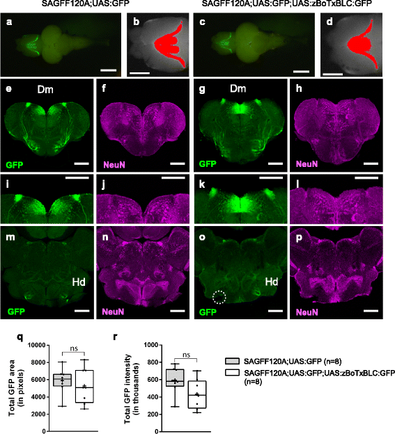
GFP expression patterns in SAGFF120A;UAS:GFP and SAGFF120A;UAS:GFP;UAS:zBoTxBLC:GFP fish. a–d GFP fluorescence in the dorsal view of the brains from SAGFF120A;UAS:GFP (~10 months old; #4 in Additional file 8: Figure S4) (a, b) and SAGFF120A;UAS:GFP;UAS:zBoTxBLC:GFP (~10 months old; #4 in Additional file 8: Figure S4) fish (c, d). Areas having more GFP intensity than background (the maximum intensity measured in the posterior part of the telencephalon) were identified by using ImageJ [57] and shown in red (b, d). c–p Immunohistochemistry using anti-GFP (green; e, g, i, k, m, o) and anti-NeuN (a neuronal marker, magenta; f, h, j, l, n, p). Coronal sections of the telencephalon (e–l) and the hypothalamus (m–p) of SAGFF120A;UAS:GFP (e, f, i, j, m, n) and SAGFF120A;UAS:GFP;UAS:zBoTxBLC:GFP (g, h, k, l, o, p) fish. i–l Magnified images of e–h. GFP-positive cells in Dm, and projections from these cells to the target area in Hd (dorsal zone of periventricular hypothalamus) were detected. A dotted circle in o indicated a broken part. Scale bars: 1 mm (a, c), 500 μm (b, d), and 200 μm (e–p). q–r Comparisons of total GFP intensity (q) and area (r) data obtained by ImageJ analysis of SAGFF120A;UAS:GFP (n = 8) and SAGFF120A;UAS:GFP;UAS:zBoTxBLC:GFP (n = 8) fish (Additional file 8: Figure S4) are plotted with Tukey box plot. Unpaired t-test with Welch’s correction was performed between these transgenic fish (ns, not significant)