
Image
|
Figure Caption
Fig. 6
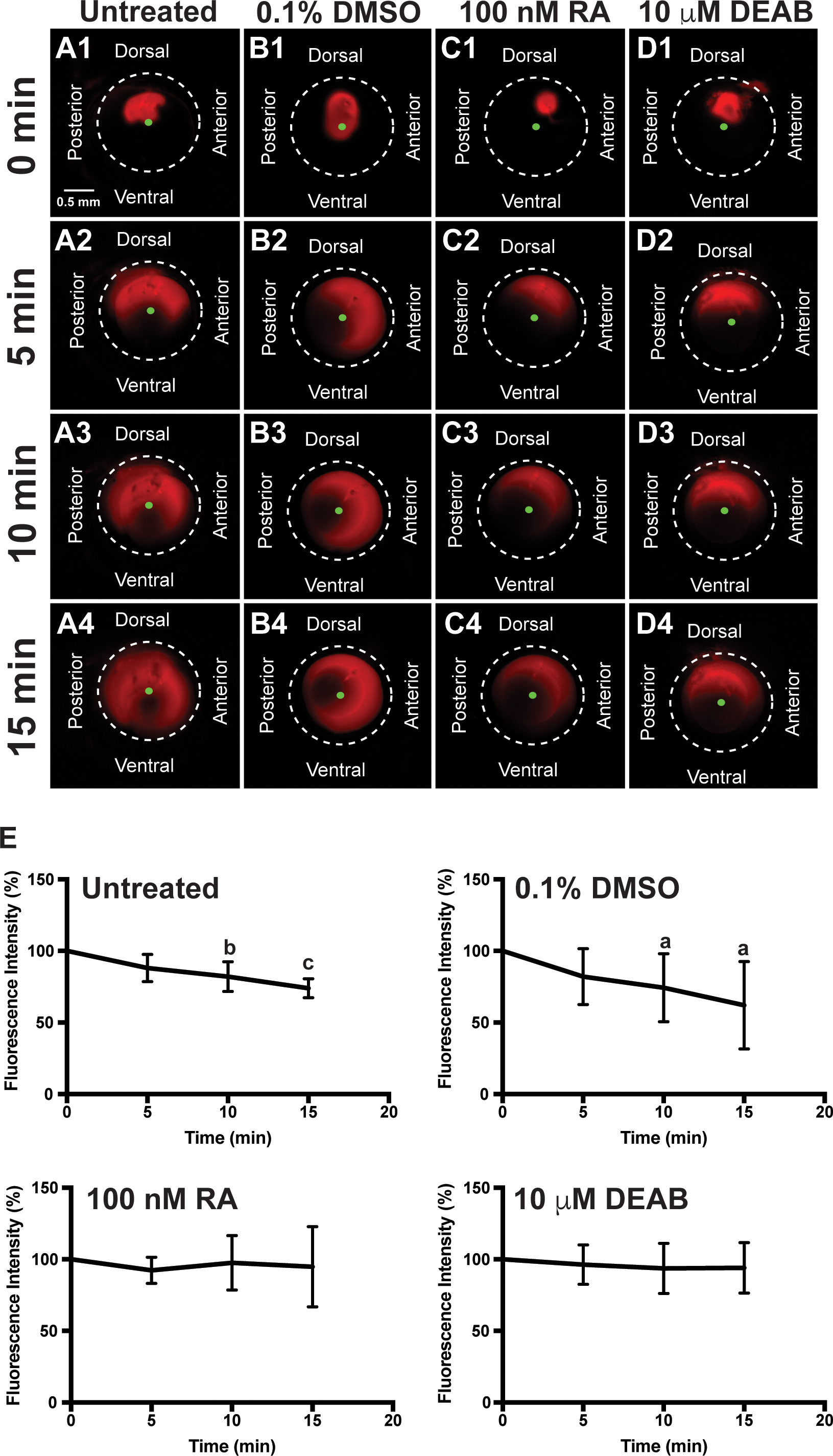
RA regulates aqueous outflow from the anterior segment of the eye. In vivo analysis of aqueous outflow showed that fluorescent Texas red dye injected into the anterior chamber diffused clockwise and counterclockwise toward the ventral iridocorneal angle over 15 minutes in untreated (A1–A4) fish. The fluorescence intensity of the dye did not change at 5 minutes (88.3% ± 9.6%), but was significantly decreased at 10 minutes (82.1% ± 10.4%) and 15 minutes (73.9% ± 6.7%) after injection (E). Similarly, the fluorescent dye diffused and exited the eye in fish treated for 2 days with 0.1% DMSO control (B1–B4). Fluorescence intensity was not changed at 5 minutes (81.8% ± 19.6%), but was significantly decreased at 10 minutes (74.3% ± 23.8%) and 15 minutes (62.0% ± 30.5%) after injection in control-injected fish (E). Fish treated with 100 nM RA for 2 days showed initial diffusion of the dye (C1), but no significant decrease in dye intensity at 5 minutes (92.3% ± 9.1%; [C2]), 10 minutes (97.6% ± 19.0%; [C3]), and 15 minutes (94.9% ± 28.1%; [C4, E]). Fish treated with 10 μM DEAB for 2 days also demonstrated initial dye diffusion (D1), but no significant decrease in dye intensity at 5 minutes (96.3% ± 13.8%; [D2]), 10 minutes (93.7% ± 17.6%; [D3]), and 15 minutes (94.1% ± 17.6%; [D4, E]).
Acknowledgments
This image is the copyrighted work of the attributed author or publisher, and
ZFIN has permission only to display this image to its users.
Additional permissions should be obtained from the applicable author or publisher of the image.
Full text @ Invest. Ophthalmol. Vis. Sci.