
Image
|
Figure Caption
Fig. 5
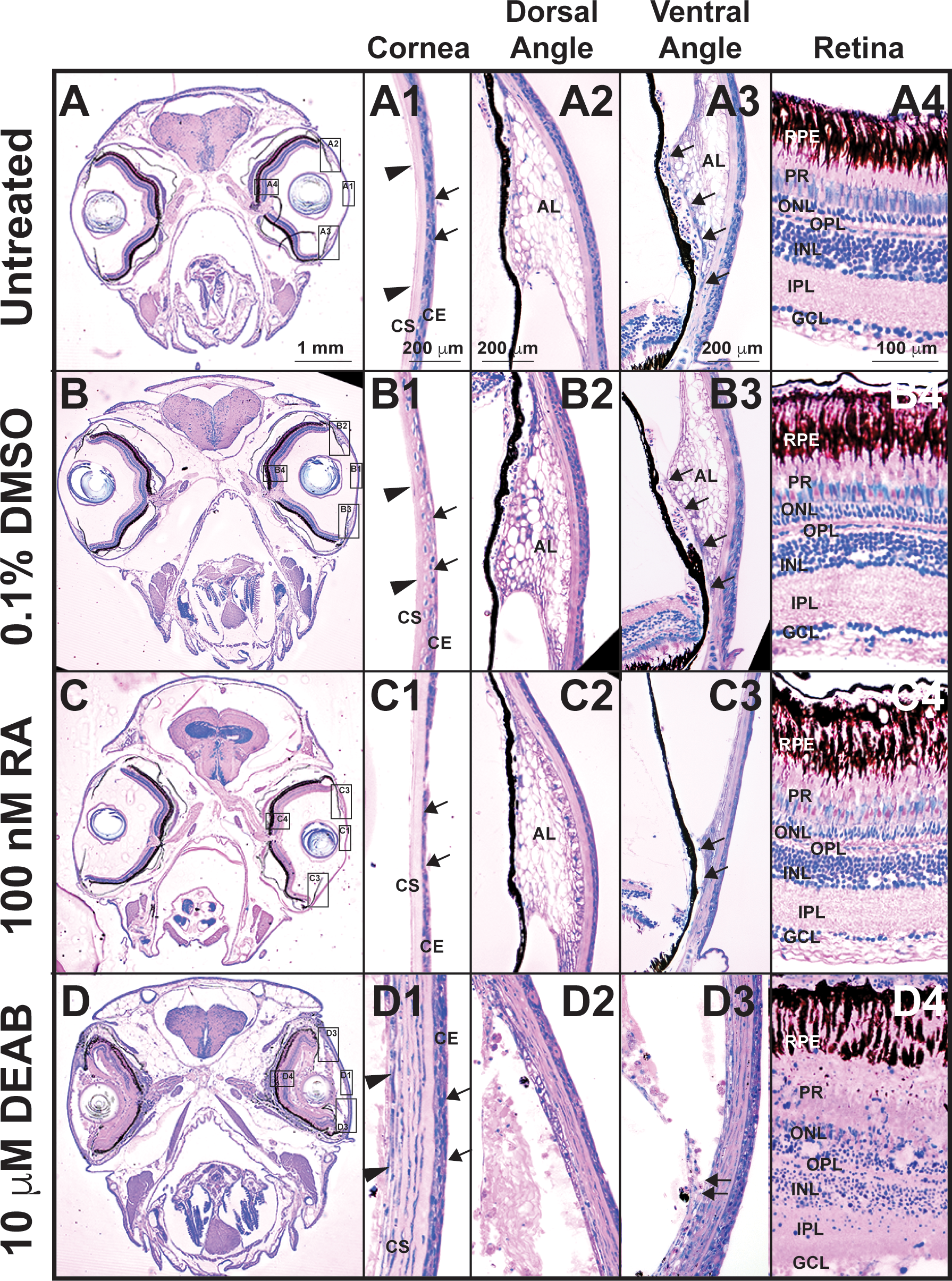
Tight control of RA levels is required for maintaining ocular structures. Methylacrylate sections of adult zebrafish treated with 100 nM RA for 5 days showed that exogenous RA decreased corneal epithelial cellularity (arrows, [C, C1]) as compared to untreated (A, A1) and DMSO control–treated (B, B1) fish. In the ventral iridocorneal angle (C3), RA also caused decreased iris stromal cellularity (C3) and loss of the webbed appearance of the AL and the ventral canalicular network (arrows, [C3]) compared to untreated (A3) and DMSO control-treated (B3) fish. The structures in the dorsal iridocorneal angle (C2) and retina (C4) were not affected by exogenous RA as compared to untreated (A2, A4) and DMSO control–treated (B2, B4) fish. Treatment with 10 μM DEAB for 5 days caused corneal stromal (arrowheads, [D, D1]) and epithelial (arrows, [D1]) edema and loss of architecture in the dorsal (D2) and ventral (D3) iridocorneal angles. DEAB also disrupted retinal structure, which included loss of PRs (D4) and ganglion cells (GCL, D4). AL, annular ligament; Co, cornea; GCL, ganglion cell layer; INL, inner nuclear layer; ONL outer nuclear layer.
Acknowledgments
This image is the copyrighted work of the attributed author or publisher, and
ZFIN has permission only to display this image to its users.
Additional permissions should be obtained from the applicable author or publisher of the image.
Full text @ Invest. Ophthalmol. Vis. Sci.