
|
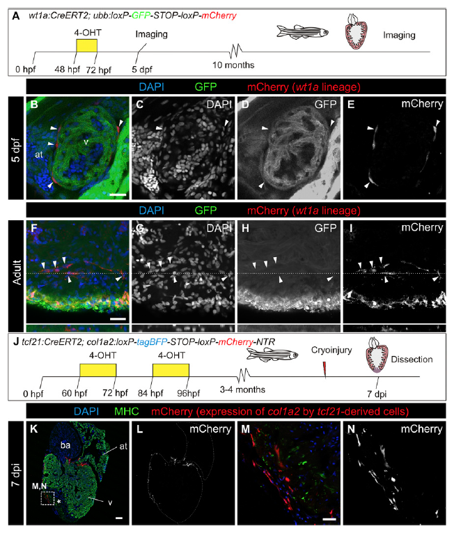
Fig. S2
Resident fibroblasts are derived from the epicardium, and cells derived from the epicardium and resident fibroblasts express col1a2:mCherry after injury. (A) Experimental scheme for tracing the fate of wt1a-derived cells. 4-OHT was administered from 48 to 72 h postfertilization (hpf). (B–E) Immunofluorescence of 5 d postfertilization (dpf) embryos with anti-GFP (green) and mCherry (red). Nuclei are counterstained with DAPI. (C–E) Single channels of the merged image shown in B. (F–I) Immunofluorescence of whole-mount adult hearts with anti-GFP (green) and mCherry (red). Nuclei are counterstained with DAPI. Orthogonal views of the plane highlighted with dotted lines are shown below. Shown are single (G–I) and merged (F) channels. (J) Experimental scheme for tracing the fate of tcf21-derived cells expressing col1a2. The tcf21:CreERT2 line was crossed with the col1a2:loxP-tagBFP-STOP-loxP-mCherry-NTR line. Upon 4-OHT administration, recombination of loxP sites leads to activation of mCherry expression under the control of a col1a2 promoter. Hearts from animals at 7 dpi were dissected and sectioned. (K–N) Immunofluorescence of the heart sections with anti-MHC (green) and mCherry (red). Nuclei are counterstained with DAPI. Asterisk indicates injured area. (Mand N) Merged and individual channels of the boxed area in K. Arrowheads mark mCherry+ cells, which express col1a2. at, atrium. [Scale bars, 25 μm (B, F, and M) and 100 μm (K).]