
|
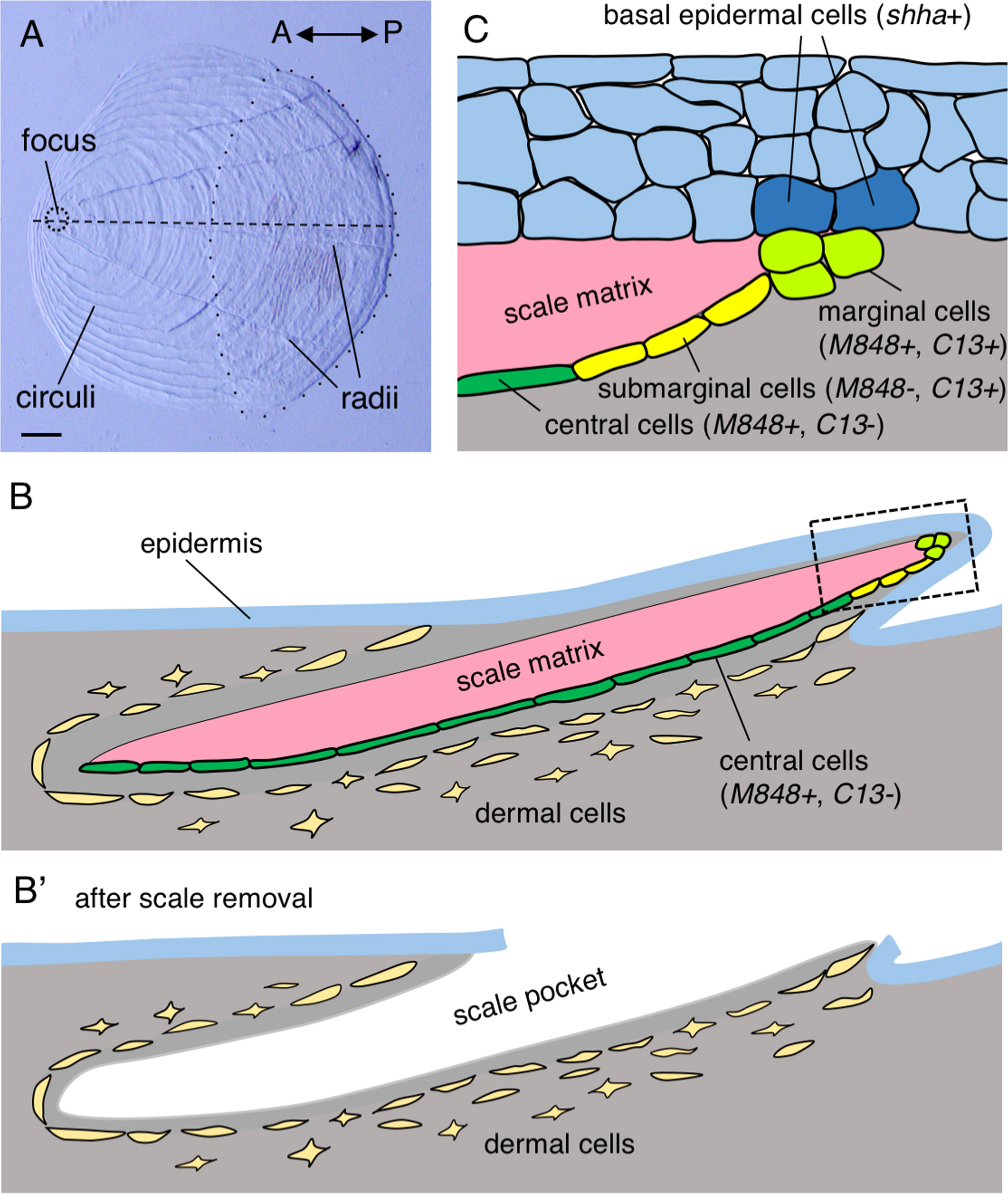
Fig. 1
Anatomy of the zebrafish scale. (A) A scale surgically removed from the trunk of an adult zebrafish. The ornamental structures (focus and circuli) are located in the anterior region, whereas radii are located in the posterior region. The posterior part of a scale (i.e. the area enclosed by the dotted line) is covered by the epidermis. (B) Schematic drawing of a scale in cross-section along the horizontal line of a scale (dashed line in (A)). The inner surface of the scale matrix is associated with central osteoblasts (dark green) and submarginal osteoblasts (yellow). The scale edge is associated with marginal osteoblasts (light green). These osteoblasts tightly attach to the matrix, and remain attached to it when a scale is removed. (B′) Dermal cells remain in the body after scale removal and contain precursor cells for the regenerating scale. (C) Structure of the skin at the posterior edge of a scale, corresponding to the boxed area in (B). Three or four layers of epidermal cells overlie the scale edge, and the basal epidermal cells make direct contact with the marginal cells. See text for detailed gene expression patterns. Scale bar, 100 µm (A).
Reprinted from Developmental Biology, 437(2), Iwasaki, M., Kuroda, J., Kawakami, K., Wada, H., Epidermal regulation of bone morphogenesis through the development and regeneration of osteoblasts in the zebrafish scale, 105-119, Copyright (2018) with permission from Elsevier. Full text @ Dev. Biol.