
|
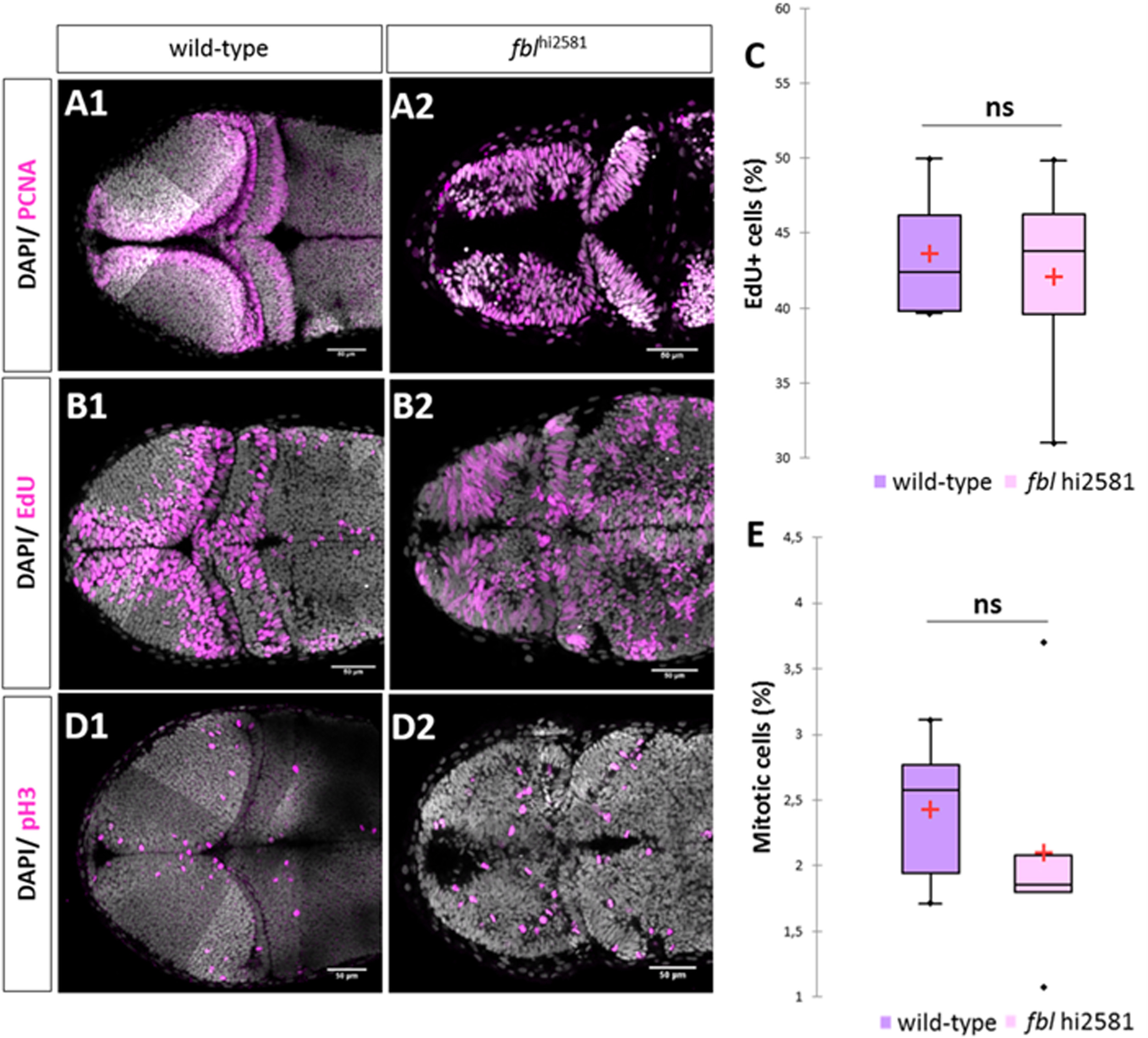
Fig. 8
In fbl mutants, the S-phase of the cell cycle is affected. (A) PCNA staining in 2dpf wild type (A1) and mutant (A2). In wild-type larvae, proliferative cells were restricted to the periphery of each tectal lobes. In fblhi2581Tg PCNA positive cells were observed in almost all tectal cells. (B) EdU incorporation experiments in 2dpf wild type (A1) and mutant (A2) embryos after two hour pulse. In wild-type embryos, EdU-positive cells are restricted to the periphery of the OT while in the fblhi2581Tg mutant embryos EdU-positive cells are spread all over the structure. (C) EdU-positive cells quantification in wild-type (purple) and mutant (pink) embryos at 2dpf. Statistical analyzes have been performed on four samples per conditions, p-value (Mann&Whitney test): 1.00. (D) pH3 staining in 2dpf wild types (E1) and mutants (E2) embryos. Similar abnormal patterns in mutants as for EdU incorporation experiments. (E) pH3-positive cells quantification in wild-type (purple) and mutant (pink) embryos at 2dpf. Statistical analyzes have been performed on four samples per conditions, p-value (Mann&Whitney test): 0.53 (E) embryos. Gray: DAPI staining, Pink: EdU, pH3 or PCNA staining. Scale bar: 50 µm. Anterior is to the left.
Reprinted from Developmental Biology, 437(1), Bouffard, S., Dambroise, E., Brombin, A., Lempereur, S., Hatin, I., Simion, M., Corre, R., Bourrat, F., Joly, J.S., Jamen, F., Fibrillarin is essential for S-phase progression and neuronal differentiation in zebrafish dorsal midbrain and retina, 1-16, Copyright (2018) with permission from Elsevier. Full text @ Dev. Biol.