
|
Fig. 5
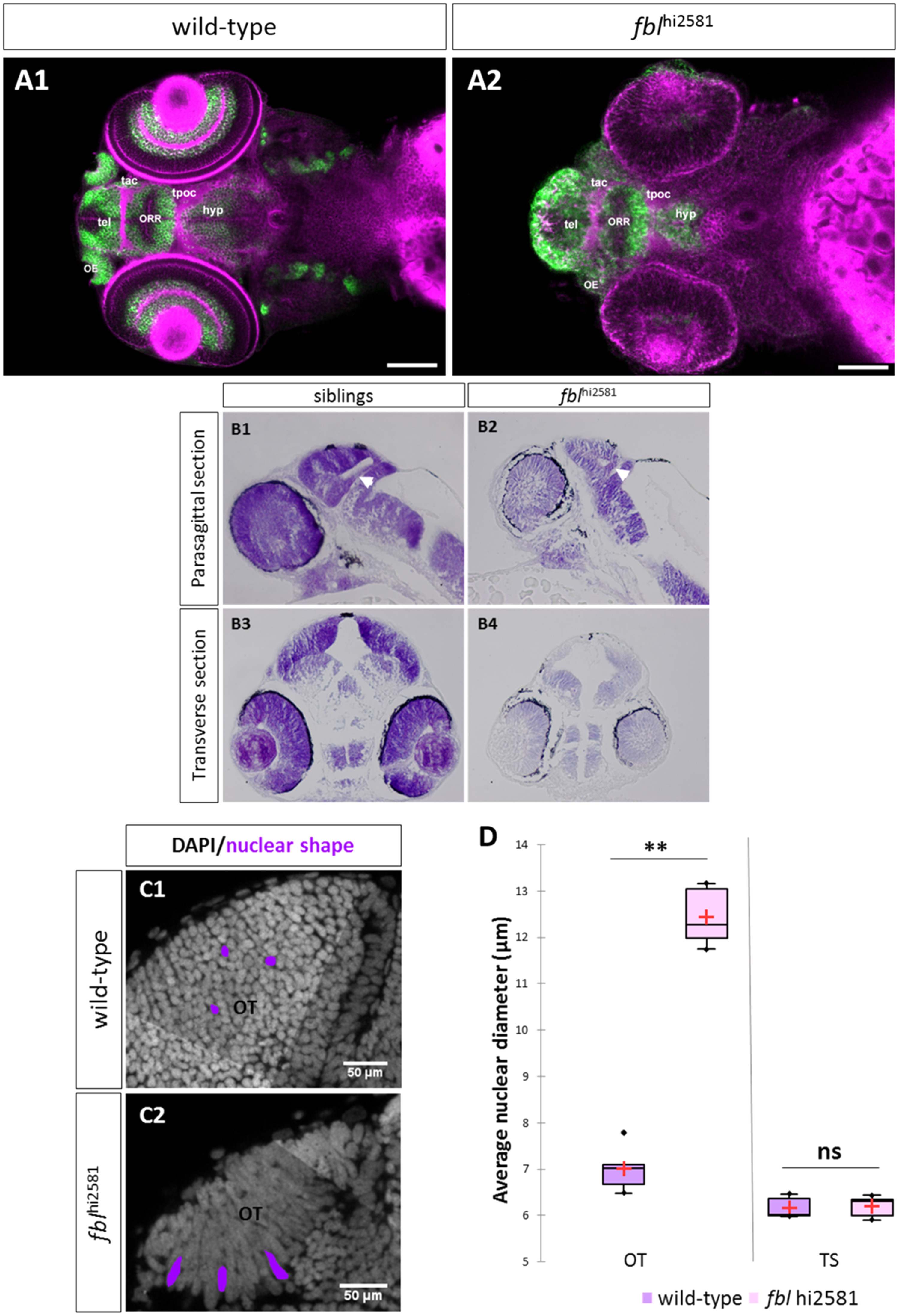
fbl mutants have specific midbrain and retina defects. (A) Horizontal optical sections of Elavl3 (marker of neural differentiation) immunolabelling and DiI staining in wild-type (A1) and mutant (A2) embryos at 3dpf. Pink: DiI labeling, Green: Elavl3 staining. Scale bars: 100 µm. Anterior is to the left. Most brain domains and axon tracts are present but have a disrupted organization in fbl mutant embryos. (B) Sagittal (A1-A2) and transverse (A3-A4) paraffin sections of wild-type (left) and fblhi2581Tg mutant (right) embryos at 2dpf. Histological analysis with cresyl violet staining revealed smaller tecta and acellular zones in the mutant embryos. The proliferation region (TMZe) is thicker in the mutant embryos (white arrow) (B2, B4) than in their siblings (B1, B3). Anterior is to the left. (C) Nuclear labeling (DAPI) in the optic tectum of 2dpf wild-type (C1) and mutant (C2) embryos at 2dpf, showing the larger nuclear diameter in the tectum of mutant larvae. Scale bar: 50 µm (D) Quantification of the nuclear diameter of wild-type (purple) and fblhi2581Tg (pink) 2dpf embryos. Nuclear diameters were measured with Fiji software. We measured the longest dimension of 50 nuclei on selected 2D images of the nuclei of 2 dpf embryos. Statistical analyses were performed on the mean diameters of nuclei from five mutant or wild-type embryos. p-value (Mann&Whitney test) OT: 0.008; p-value TS: 1.000. Hyp: hypothalamus, OE: olfactory epithelium, ORR: optic recess region OT: optic tectum, tac: tract of the anterior commissure, tel: telencephalon, tpoc: tract of the post-optic commissure, TS: torus semicircularis.
Reprinted from Developmental Biology, 437(1), Bouffard, S., Dambroise, E., Brombin, A., Lempereur, S., Hatin, I., Simion, M., Corre, R., Bourrat, F., Joly, J.S., Jamen, F., Fibrillarin is essential for S-phase progression and neuronal differentiation in zebrafish dorsal midbrain and retina, 1-16, Copyright (2018) with permission from Elsevier. Full text @ Dev. Biol.