
|
Fig. 1
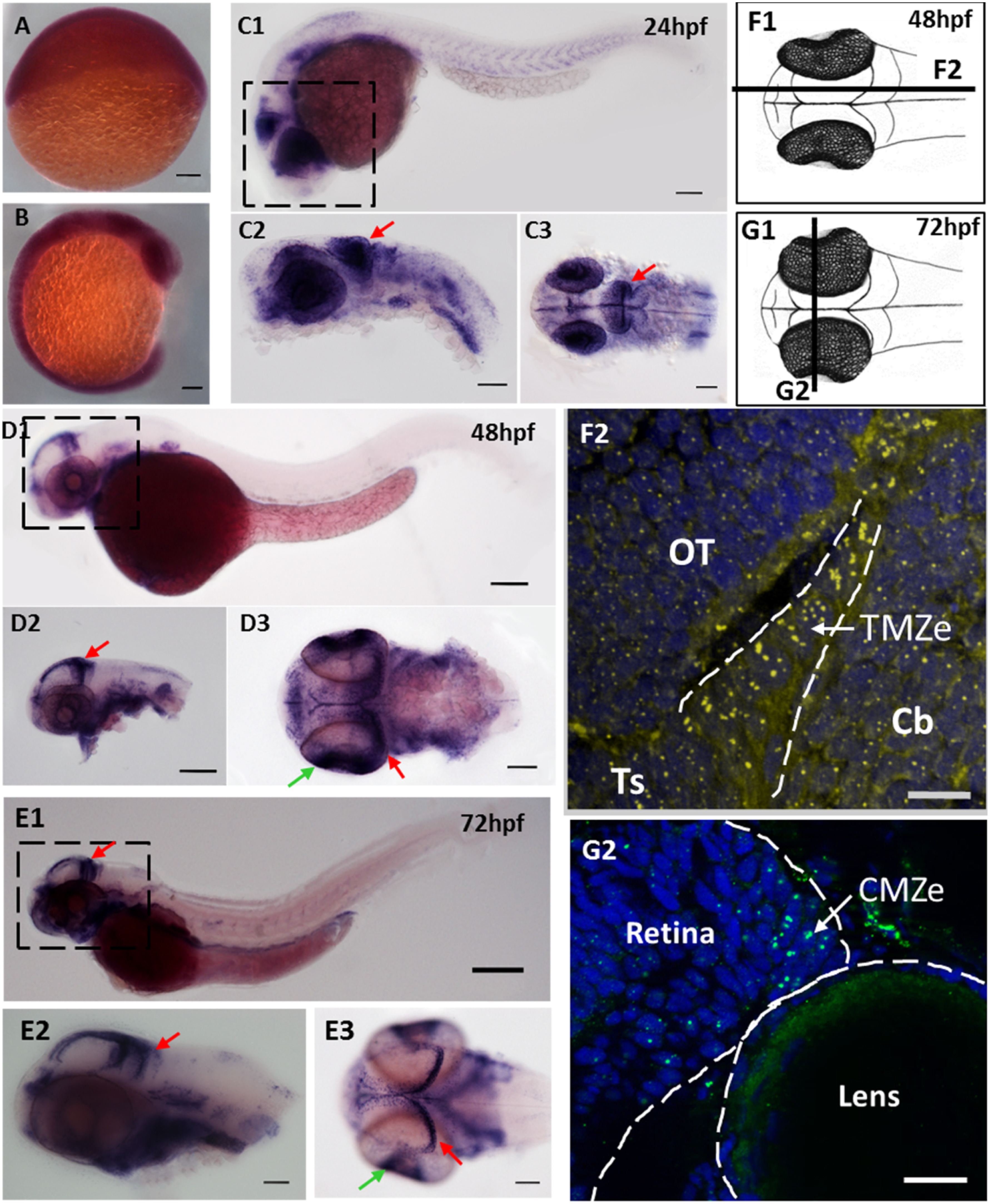
fbl transcripts and Fbl protein are preferentially expressed in neural progenitors during zebrafish development. (A-E)In situ hybridization showing the progressive restriction of fbl expression during the development of zebrafish embryos (A)fbl is ubiquitously expressed at 6 hpf. (B). fbl expression begins to be restricted to highly proliferative regions (eyes, midbrain, and somites) during neurulation (C) At 24 hpf, fbl transcripts are abundant in the optic tectum (red arrows) and the retina. C1-C2: lateral views, C3: dorsal view (D-E) At 2 dpf (D) and 3 dpf (E), fbl is preferentially expressed in the neuroepithelial progenitors of the TMZe at the periphery of the OT (red arrows), and in the external edge of the ciliary marginal zone (CMZe) of the retina (green arrows). Additional expression can be detected in the digestive system D1-D2 and E1-E2: lateral views, D3 and E3: dorsal views. Scale bars: 100 µm. Anterior is to the left. (F) F1: Drawing of a 2dpf zebrafish embryo in dorsal view. The dark line corresponds to the sagittal section represented in F2. F2: Immunostaining showing Fbl protein on a sagittal section of a 2 dpf embryo. The Fbl protein, which is localized in the nucleoli, is present in all cells (yellow dots) but the punctate domains of expression are larger in TMZe neuroepithelial progenitors (surrounded by white dashed lines) than in other cells in the optic tectum (OT), cerebellum or torus semicircularis. (G) G1: Drawing of a 3dpf zebrafish embryo in dorsal view. The dark line corresponds to the transverse section represented in G2. G2: Immunostaining showing Fbl protein on a transvere section of a 3dpf embryo. The Fbl protein accumulates at the periphery of the retina where neuroepithelial cells are located (CMZe). Scale bars: 25 µm. Cb: cerebellum; CMZe: external edge of the ciliary marginal zone; OT: optic tectum; Ts: torus semicircularis;; TMZe: external edge of the tectal marginal zone.
Reprinted from Developmental Biology, 437(1), Bouffard, S., Dambroise, E., Brombin, A., Lempereur, S., Hatin, I., Simion, M., Corre, R., Bourrat, F., Joly, J.S., Jamen, F., Fibrillarin is essential for S-phase progression and neuronal differentiation in zebrafish dorsal midbrain and retina, 1-16, Copyright (2018) with permission from Elsevier. Full text @ Dev. Biol.