
|
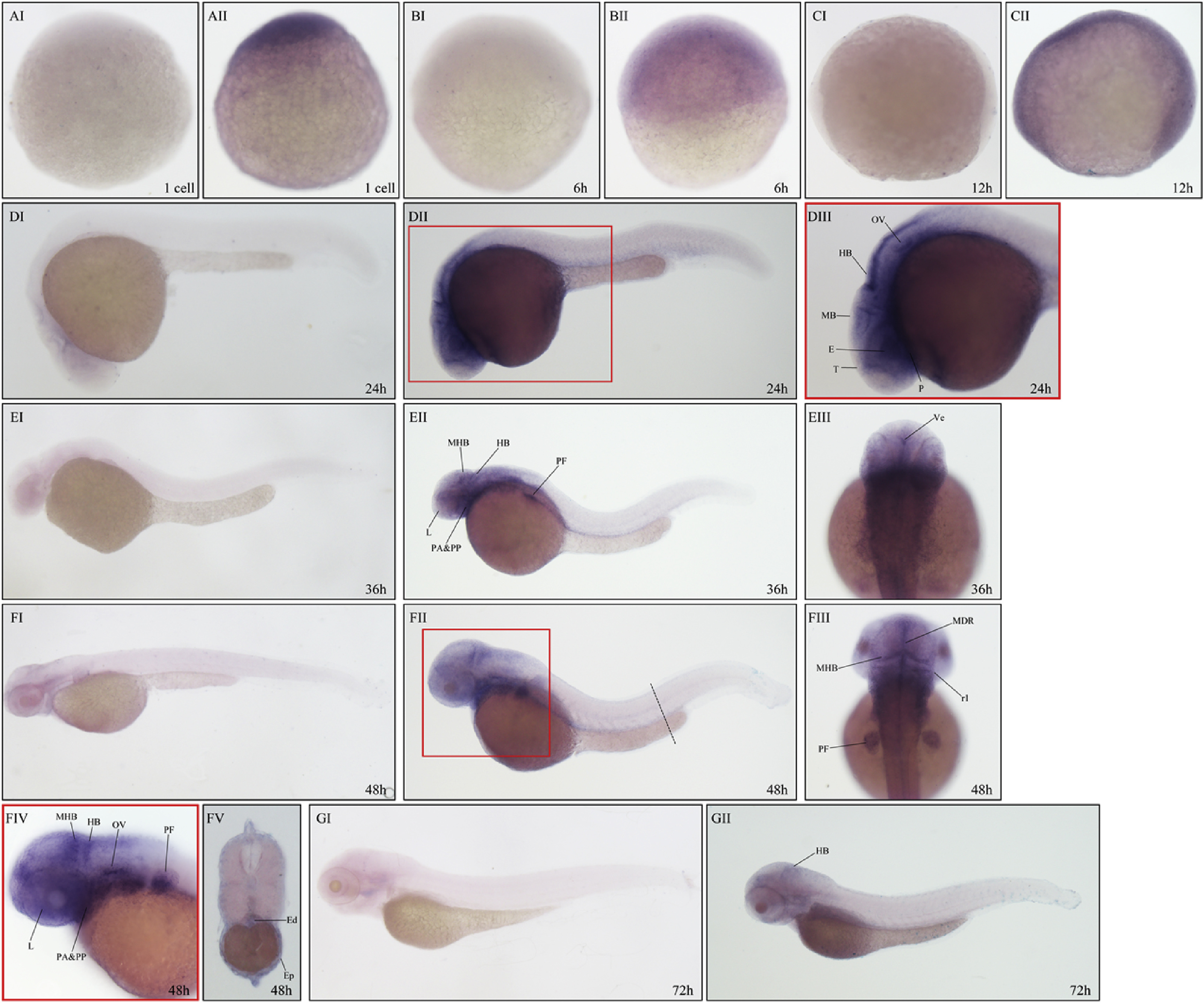
Fig. 2
Expression of vgll4a in zebrafish embryos analyzed by WISH.
Expression of vgll4a at one cell stage (AII), 6 hpf (BII), 12 hpf (CII), 24 hpf (DII-DIII), 36 hpf (EII-EIII), 48 hpf (FII-FV) and 72 hpf (GII). Embryos incubated with vgll4a sense probe are shown as negative controls (AI, BI, CI, DI, EI, FI, GI). Embryos are shown in lateral view with anterior to the left (DI-EII, FI, FII, FIV, GI, GII) or dorsal view with anterior to the top (EIII, FIII). Box areas in (DII) and (FII) was shown enlarged in (DIII) and (FIV) respectively. Dotted line in (FII) indicates approximate orientation of cross section image showed in (FV). T, telencephalon; E, eyes; MB, midbrain; HB, hindbrain; OV, otic vesicle; P, pharynx; L, lens; PF, pectoral fin; PA, pharyngeal arches; PP, pharyngeal pouches; Ve, ventricle; MHB, midbrain–hindbrain boundary; MDR midline of diencephalic roof; r1, rhombomere 1; Ed, endoderm; Ep, epidermis.
Reprinted from Gene expression patterns : GEP, 28, Xue, C., Wang, H.H., Zhu, J., Zhou, J., The expression patterns of vestigial like family member 4 genes in zebrafish embryogenesis, 34-41, Copyright (2018) with permission from Elsevier. Full text @ Gene Expr. Patterns