
|
Fig. 4
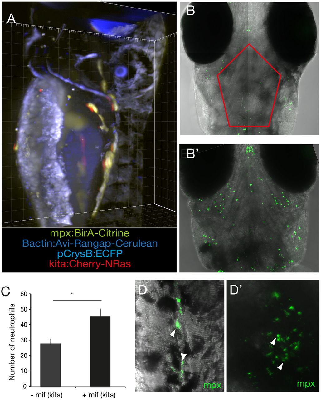
mCherry-NRasK61Q-activated melanocytes are immunogenic. (A) Larvae at 5 dpi harbouring all four alleles (blue eye, blue nuclei, green neutrophils and red melanocytes) as captured on a Zeiss Z1 Light Sheet microscope. kita:LexPR;LexOP:NRasQ61K were crossed to mpx:BirA;bactin:Avi-Rangap, and mCherry-NRas activated by addition of mifepristone. (B) The melanocyte-specific oncogene NRasK61Q was activated by addition of mifepristone to the E3 medium in Tg(kita:LexPR-Cerulean;pCrysβ:ECFP-LexOP:mCherry-NRasQ61K)ox129 embryos at 24 hpf. At 5 dpi, composite confocal images show neutrophils (green) as detected with an anti-mpx antibody in stage-matched controls (−mif) (B) and embryos where the oncogene had been activated (+mif) (B′). Images are a maximum intensity projection of 11 z-stack projections captured from the surface of the embryo to a depth of 100 μm. (C) Quantification of the number of neutrophils in the region of cranial melanocytes in the outlined area in B in control larvae (−mif, n=8) and following oncogene activation (+mif, n=8). Graph shows means±s.e.m. Statistical significance was determined by two-tailed unpaired Student's t-test with Welch's correction. **P<0.01. (D) High-resolution image of neutrophils in the tail (D) and head (D′) of 5-dpf zebrafish larvae following oncogene activation at 24 hpf, as shown by the arrowheads.