
|
Fig. 5
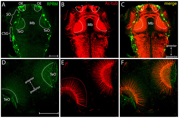
Expression of RRPM in zebrafish tectum opticum. Whole-mount immunofluorescence showing RPRM staining in a representative zebrafish embryo at 3dpf. (A–C) Dorsal view with anterior to the top, posterior to the bottom (double arrows in C). Expression of RPRM is shown in green and Ac-tub is shown in red. Positive labeling is observed in the OE, supraorbital neuromasts (SO, brackets), cranial sensory ganglia (CSG, brackets) and TeO (dotted white areas in A–C). (D–F) Magnification of TeO area with anterior to the upper-left and posterior to the bottom-right (double arrows in D), showing optic neuropil area labeled bilaterally (dotted white areas in D) as well as projections extending radially toward the midline (red dotted area in E). (F) Merged image for (D,E). (A–F) All the panels represent z-stack projections. Scale Bars in (A,D): 100 μm.