
|
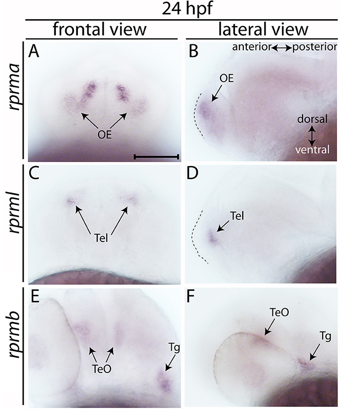
Fig. 1
Expression of RPRM mRNA at 24 h post-fertilization (hpf). (A–E) The expression patterns of rprma/b and rprml were visualized by whole-mount in situ hybridization (WISH) during zebrafish early neuronal development. (A,C,E) Frontal views of the embryos heads, with dorsal to the top and ventral to the bottom (double arrow in B). (B,D,F) Lateral views of the embryos head with anterior to the left and posterior to the right (double arrow in B). (B,D) dashed areas represent the most anterior part of the embryo's head. At 24 hpf (A,B) rprma, (C,D) rprml, and (E,F) rprmb transcripts are located in neuronal cell populations such as: (A,B) olfactory placode (OP, black arrows), (C,D) telencephalon (Tel, black arrows), and (E,F) tectum opticum (TeO) and trigeminal ganglia (Tg), respectively. Scale bar in (A): 100 μm.