
|
Fig. S3
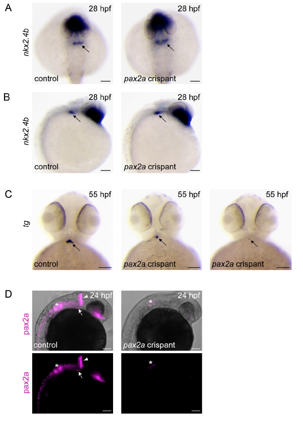
Thyroid anlage specification is not perturbed in pax2a crispants. (A,B) Whole-mount in situ hybridization for the early thyroid marker nkx2.4b in non-injected control embryos and pax2a crispants. Thyroidal nkx2.4b expression (arrow) was not different between experimental groups at 28 hpf. Panel A shows dorsal views of stained specimen (anterior is to the top). Panel B shows lateral views (anterior is to the right). (C) Loss of thyroid marker expression in later stage pax2a crispants. Whole-mount in situ hybridization for the thyroid differentiation marker tg in non-injected control embryos and pax2a crispants at 55 hpf. In contrast to the strong tg staining in the compact and slightly ovoid thyroid primordium of control embryos, half of the pax2a crispants showed either a complete absence of detectable tg staining (data not shown) or presented thyroid primordia of greatly reduced size. Ventral views are shown (anterior is to the top). (D) pax2a crispants display strongly reduced immunoreactivity to a pax2a antibody. Whole-mount immunofluorescence staining of pax2a (magenta) was performed on non-injected control embryos and pax2a crispants at 24 hpf using a pax2a antibody directed against an epitope located C-terminal to the sgRNA target site. Approximately half of the pax2a crispants (53.7%; N=79/147) displayed a strongly reduced pax2a immunofluorescence signal. Lateral views are shown (anterior is to the right). Arrowheads: mid-hindbrain boundary; arrows: thyroid anlage; asterisks: otic vesicle. Scale bars: 100 μm.