
|
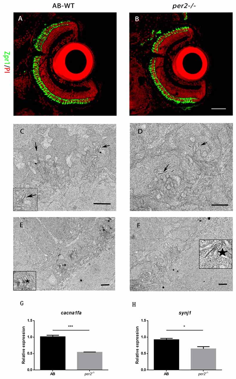
Fig. 3
per2 mutant zebrafish larvae exhibited normal retinal morphology and abnormal photoreceptor ribbon synapses. (A,B) AB-WT and per2−/− zebrafish larval retinas revealed relatively normal retinal morphology by IHC. Images are LSCM presentations. Larvae at 5 dpf were stained with zpr1 (green) and propidium iodide (PI; red), which label double cones and nuclei (n = 6 animals per genotype). Scale bar = 40 μm. (C–F) per2 mutant zebrafish larvae exhibited abnormal ribbon synapses. (C) In the AB-WT retina, synaptic ribbons (arrows) are associated with the presynaptic membrane via an arciform density (asterisks). (D) In the per2−/− retina, synaptic ribbons in most of the pedicles appear to be floating in the cytoplasm and are unassociated with an arciform density and the presynaptic membrane. AB-WT (E) had relatively more normal ribbon synapses (asterisks) than those of the per2 mutant (F; n = 3 retina per genotype). Scale bar = 1 μm. (G,H) Expression of cacnf1a and syjn1 was reduced in mutant compared with WT retinas (5 dpf, n = 10 animals per genotype, *P < 0.05 and ***P < 0.001, unpaired two-tailed t-tests). Data represent the mean ± SEM.