
|
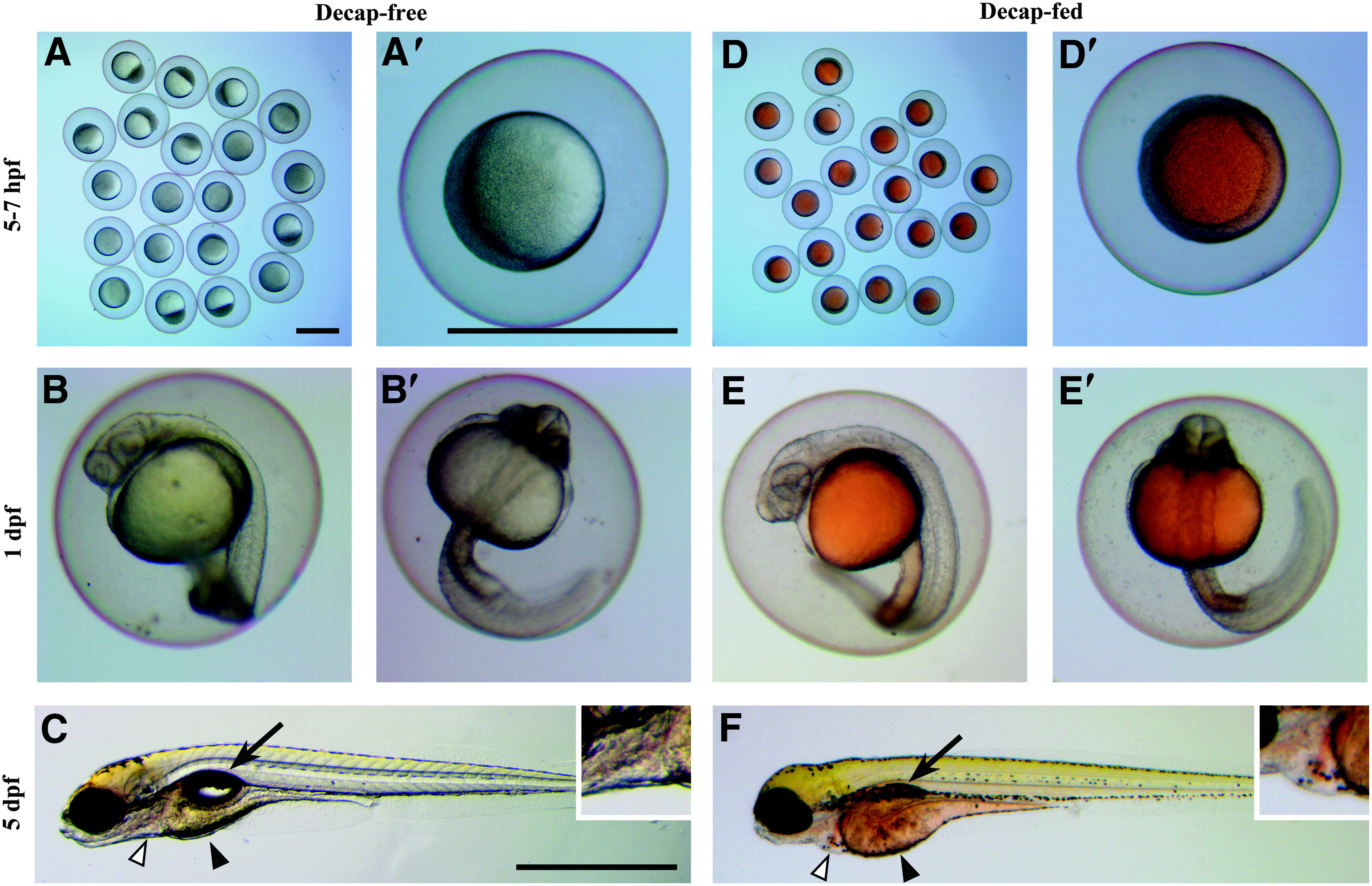
Fig. 1
Comparison of yolk coloration and morphological deformities between progeny of adults that were fed decapsulated brine shrimp cysts (Decap-fed) and progeny of adults that were not fed decapsulated brine shrimp cysts (Decap-free). (A–C) Progeny of Decap-free adults exhibited clear or pale yellow yolks and normal morphological features. Low (A) and high (A′) magnification of 5–7 hpf embryos. Lateral (B) and ventral (B′) views of 1 dpf embryos. (C) Lateral view of a 5 dpf larva with normal inflated swim bladder (arrow), cardiac sac (white arrowhead and inset, enlarged 200%), and pale-yellow yolk (black arrowhead). (D–F) Progeny of Decap-fed adults exhibited orange coloration in yolks and abnormal morphological features. Low (D) and high (D′) magnification of 5–7 hpf embryos with orange-colored yolks. Lateral (E) and ventral (E′) views of 1 dpf embryos, which continued to exhibit orange coloration in the yolk and yolk extension. (F) Lateral view of a 5 dpf larva that appeared developmentally delayed with an uninflated swim bladder (arrow), cardiac edema (white arrowhead and inset, enlarged 200%), and an enlarged orange-colored yolk (black arrowhead). Scale bars = 1 mm (A applies to A and D; A′ applies to A′, B, B′, D′, E, and E′; C applies to C and F). Hours postfertilization, hpf; days postfertilization, dpf.