
|
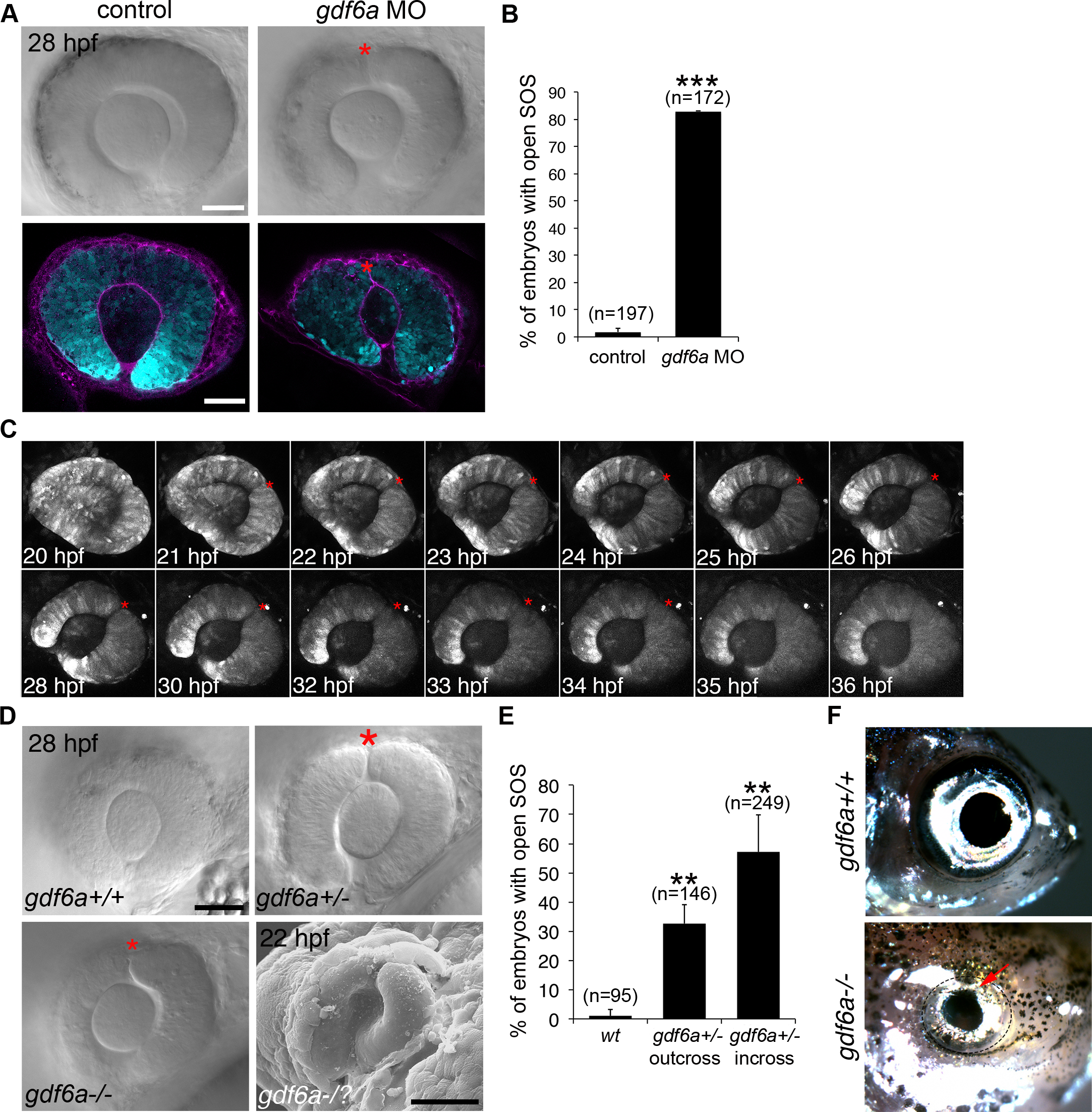
Fig. 4
The role of Gdf6a signaling in superior ocular sulcus morphogenesis.
(A) Delayed SOS closure caused by Gdf6a knockdown. Tg(rx3:GFP) zebrafish eyes (cyan) from uninjected and Gdf6a morpholino-injected embryos shown as DIC images of live embryos and single optical slices following anti-Laminin antibody staining (magenta). SOS marked by red asterisk. (B) Quantification of embryos with delayed sulcus closure, as assessed at 28 hpf. (C) Time series of maximum projection confocal images of a Tg(rx3:GFP) embryo injected with gdf6a morpholino. (D) DIC images of wildtype, gdf6a+/- and gdf6a-/- eyes (SOS marked by red asterisk). Bottom right panel shows SEM image of a Gdf6a-deficient eye with a pronounced sulcus. (E) Quantification of gdf6a-/- mutants (or siblings) with delayed SOS closure. (F) Adult wildtype zebrafish (top panel) showing normal eye morphology and a gdf6a-/- zebrafish (bottom panel) with superior coloboma (red arrow). N = 3 experiments for graphs in B and E. n = number of embryos. Data are means ± SEM. Statistics in B is a two-tailed t test, and in E is one-way ANOVA with Tukey’s test: **P<0.01, *** P<0.001. Scale bars are 50 μm.