
Image
|
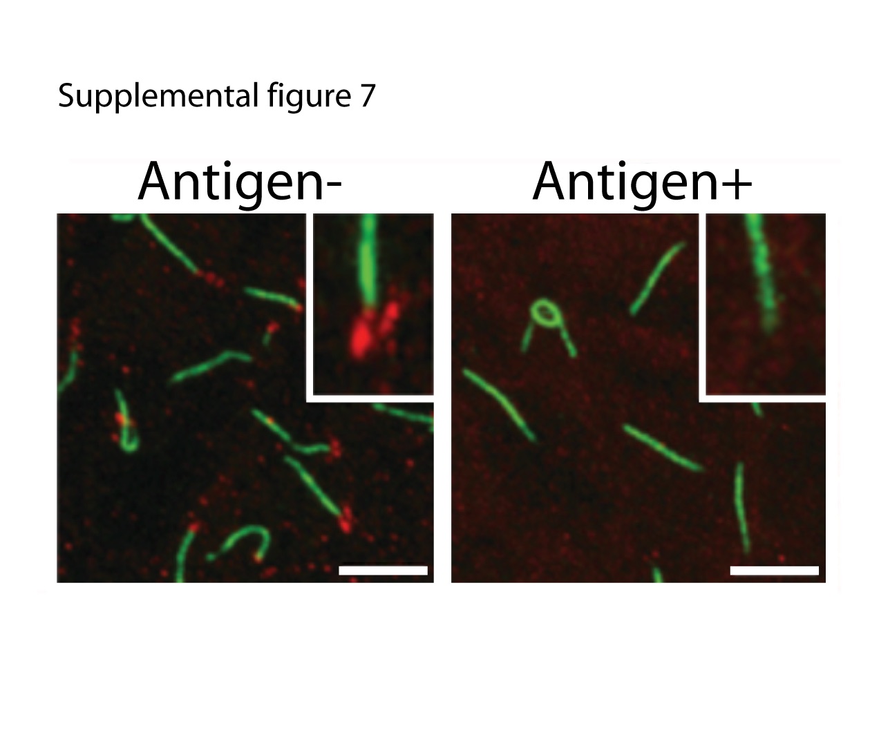
Figure Caption
Fig. S7 Anti-Cep290 antibody validation. In a 10 somite stage zebrafish embryo, the Cep290CT antibody (red) stain punctate structures at the base of Kuppfer's vesicle cilia (anti-acetylated tubulin in green). Staining is lost when the antibodies are preincubated with antigen, demonstrating antibody specificity. Inset: magnified view of a single basal body region. Scale bar is 5 µm.
Acknowledgments
This image is the copyrighted work of the attributed author or publisher, and
ZFIN has permission only to display this image to its users.
Additional permissions should be obtained from the applicable author or publisher of the image.
Full text @ Cilia