
Image
|
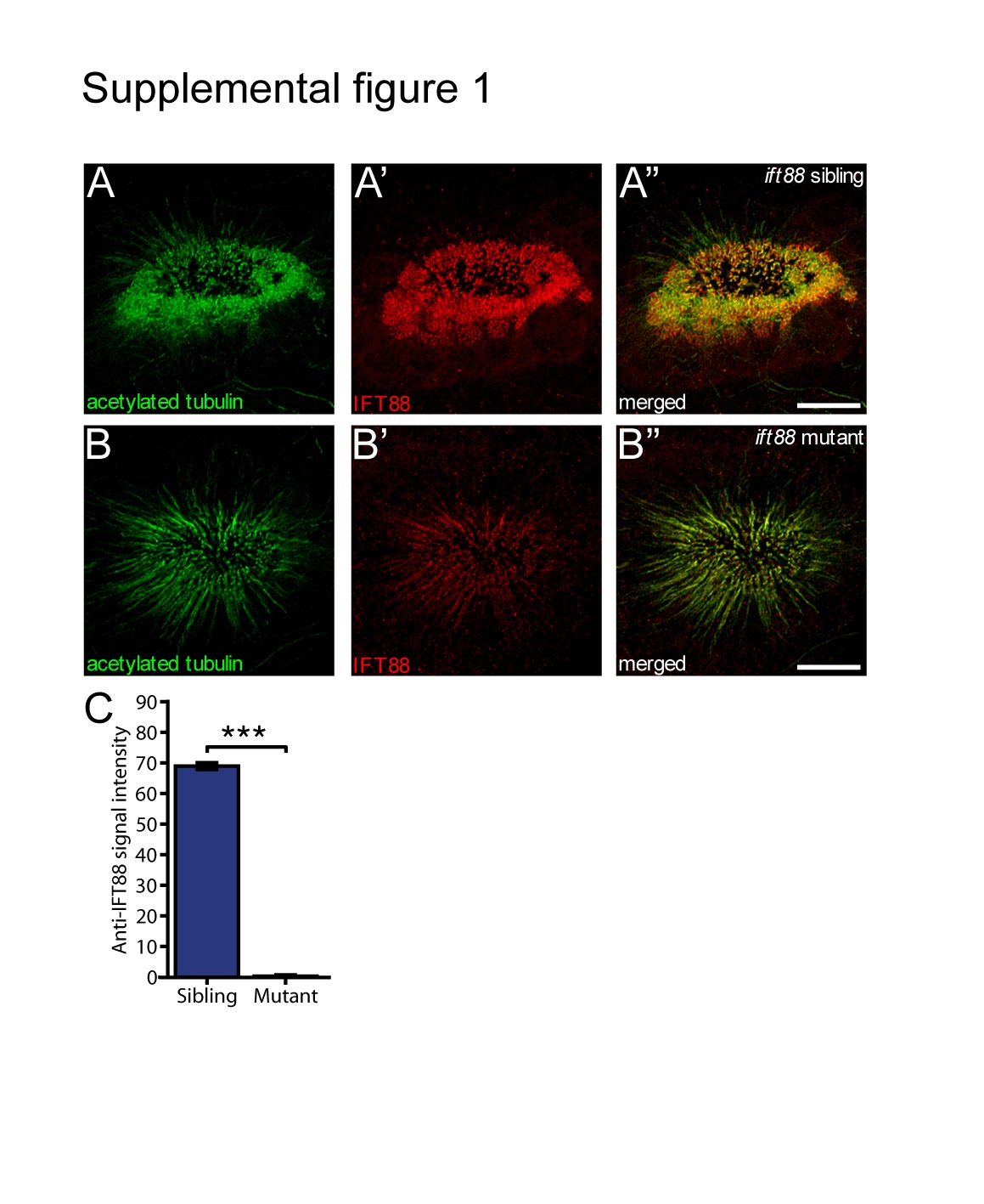
Figure Caption
Fig. S1
Severe reduction of IFT88 protein (anti-IFT88 antibody in red) expression is present in the cilia of the OE (marked by anti-acetylated tubulin staining) of the ift88 mutant (B) compared to the ift88 wildtype sibling (A). (C) Quantification of the signal in the red (IFT88) channel demonstrated a 99% decrease in intensity of the red staining. (N=3 fish per condition, P=5.5E-7, Student's t-test). Bars represent mean and SEM. Scale bar is 10 µm.
Acknowledgments
This image is the copyrighted work of the attributed author or publisher, and
ZFIN has permission only to display this image to its users.
Additional permissions should be obtained from the applicable author or publisher of the image.
Full text @ Cilia