
|
Fig. 1
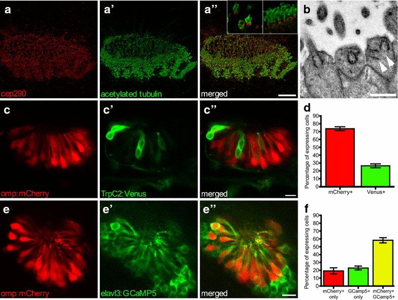
Olfactory epithelium (OE) in 2.5-dpf zebrafish. Multi-ciliated motile cilia are present at the border, ciliated OSNs in the centre; maximum intensity projection. a Basal bodies stained with anti-cep290 (red), a’ cilia stained with anti-acetylated tubulin (green), a” merged image. Inset in a” closeup of sensory (left) and motile (right) cilia showing basal body staining of anti-Cep290. b EM image of dendritic knob of ciliated zebrafish OSNs containing multiple basal bodies (arrowheads). c Tg(omp:mCherry,trpc2:Venus) zebrafish with ciliated Omp-positive OSNs (red) and c’ microvillus TrpC2-positive OSNs (green), c” merged image; single confocal section. d Quantification of C, N = 5 fish. e Tg(omp:mCherry,elavl3:GCaMP5) zebrafish show overlap between ciliated Omp-positive OSNs (red) and e’ GCaMP5-positive OSNs (green) OSNs, e” merged single confocal section. f Quantification of E, N = 6 fish. Bars represent mean and SEM. Scale bar is 10 µm, except b bar is 1 µm