
|
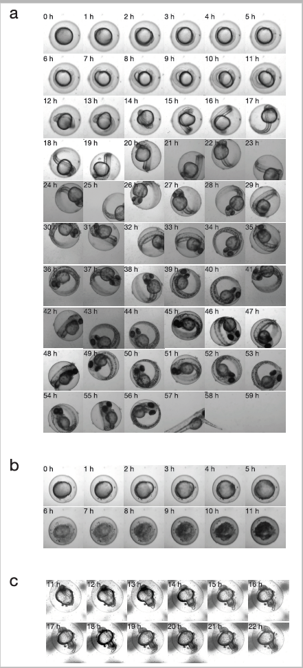
Fig. S1
Developmental dynamics of zebrafish embryos showing normal development and examples of embryonic death. After UV irradiation, embryonic survival was tracked by time-lapse imaging in 1-h intervals until the hatch period (at least 60 h). The elapsed time from the start of recording is indicated in the upper left corner in each panel. (a) Entire time-lapse sequences of the typical normal development of a zebrafish embryo. (b, c) Examples of embryonic death; in these cases, we determined that embryonic death occurred at 6 h (b) and 19 h (c), respectively. In both frames, critical deformation and cessation of development were observed.