
Image
|
Figure Caption
Fig. S3
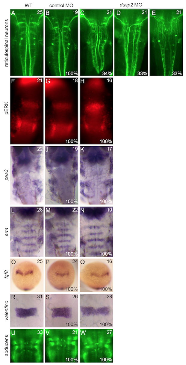
Additional neuronal and patterning markers examined in dusp2 morphants. Wildtype, control MO-injected, and dusp2 MO-injected embryos were analyzed by in situ hybridization for the expression of pea3, erm, fgf8, and valentino and by immunostaining to visualize the reticulospinal neurons, pERK, and the abducens motor neurons.
Acknowledgments
This image is the copyrighted work of the attributed author or publisher, and
ZFIN has permission only to display this image to its users.
Additional permissions should be obtained from the applicable author or publisher of the image.
Full text @ BMC Dev. Biol.