
|
Fig. 1
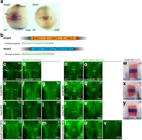
Knockdown of dusp2 and dusp6 via MO yields a hindbrain phenotype. a 12hpf wildtype embryos were assayed by in situ hybridization for expression of krox20 (red stain) and dusp6 (blue stain in left panel) or dusp2 (blue stain in right panel). b Schematic of genomic sequence for dusp6 and dusp2. Red vertical lines indicate CRISPR target sites and green vertical lines indicate MO target sites. c-v 48hpf wildtype (c, n), control MO-injected (d, o), dusp6 MO-injected (e-g, p-q), dusp2 MO-injected (h-j, r-s), and dusp6 + dusp2 MO-injected (k-m, t-v) embryos were assayed by immunostaining for differentiation of Mauthner neurons (3A10 staining in c-m) and facial motor neurons (Islet1/2 staining in n-v). w-y 18hpf wildtype (w), control MO-injected (x) and dusp2 MO-injected (y) embryos were assayed by in situ hybridization for expression of krox20 (red stain) and hoxb1a (blue stain). Numbers in top right corner of each panel indicate the total number of embryos assayed for that condition. Numbers in bottom right corner indicate percent of embryos with the phenotype shown. All embryos are in dorsal view with anterior to the top. Embryos in (a) are whole-mounts, while embryos in (c-y) are flat-mounted and show only the central hindbrain region