
Image
|
Figure Caption
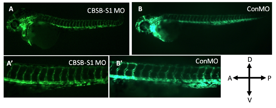
Fig. S3
Lack of vascular phenotype in cbsb (~50 hpf) morphants. The Tg (flk:EGFP) fish used in the figure 5 experiment carried a VEGFR2 promoter (FLK) driving enhanced green fluorescent protein (EGFP) in the vasculature. A and A’ are cbsb splice MO1 (CBSB-S1) MOinjected fish while B and B’ are control (Con) MO-injected fish. A’ and B’ are high magnification images of the trunk vasculature. In the figure panels, the embryo orientation is left is anterior (A) and right is posterior (P) while top is dorsal (D) and bottom is ventral (V).
Acknowledgments
This image is the copyrighted work of the attributed author or publisher, and
ZFIN has permission only to display this image to its users.
Additional permissions should be obtained from the applicable author or publisher of the image.
Full text @ Front Cell Dev Biol