Image
Figure Caption
Fig. 2-S1
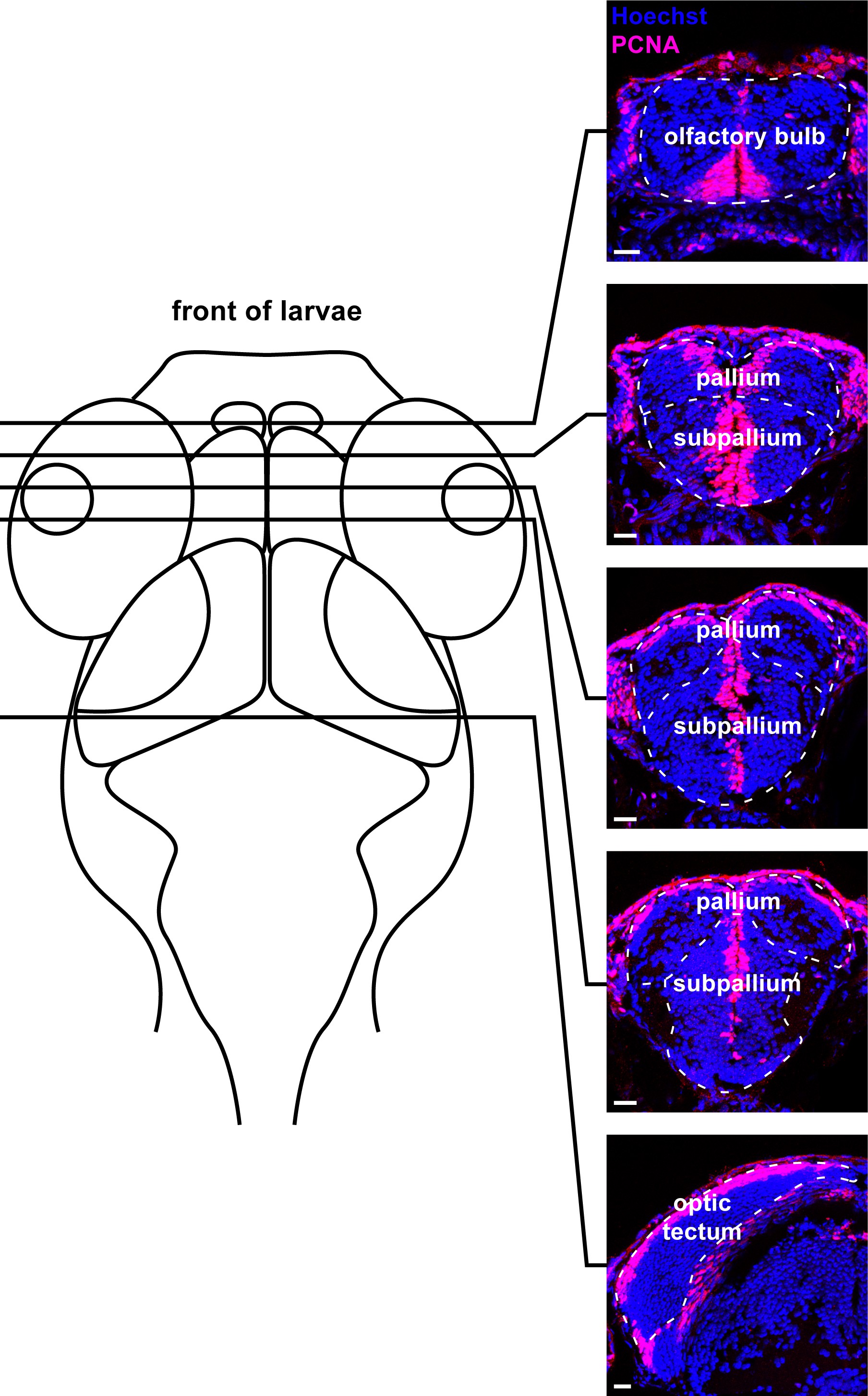
Example traces of brain regions sampled through coronal sections in the larval zebrafish brain.
Micrographs (20 µm thickness) with example boundaries traced for the olfactory bulb, pallium, subpallium, and optic tectum (white dotted line) along with their approximate rostrocaudal position on a schematic of a dorsal view of the larval zebrafish head. Scale bars = 20 µm.
Acknowledgments
This image is the copyrighted work of the attributed author or publisher, and
ZFIN has permission only to display this image to its users.
Additional permissions should be obtained from the applicable author or publisher of the image.
Full text @ Elife

