
|
Fig. 2
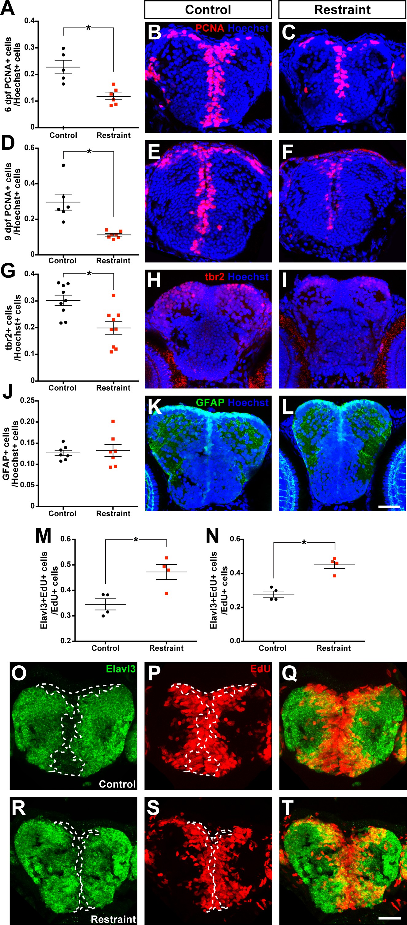
Movement restraint reduces cell proliferation in the larval forebrain.
By 6 dpf, movement restraint reduces the proportion of PCNA+ cells in the forebrain (A-C; control n = 5, restraint n = 6). This reduction in PCNA +cells is maintained when movement restraint is continued until 9 dpf (D-F; control n = 6, restraint n = 7). Movement restraint until 9 dpf also reduces tbr2+ cells in the pallium (G-I; n = 9) without affecting the number of GFAP+ radial glia stem cells in the pallium (J-L; n = 7; scale bar for micrographs in B-L = 30 µm). Following a 24 hr pulse with EdU starting on 5 dpf, fewer EdU+ cells in the subpallium (M) and pallium (N; n = 4) co-label for the neuronal fate marker Elavl3 in controls (O–Q) compared to movement restrained larvae (R-T; scale bar = 20 µm). White dotted lines mark the boundaries of Elavl3+ expression to highlight the increased overlap between EdU+ cell cohorts and Elavl3+ in restrained larvae. *p<0.05. Data are represented as mean ± SEM.