
|
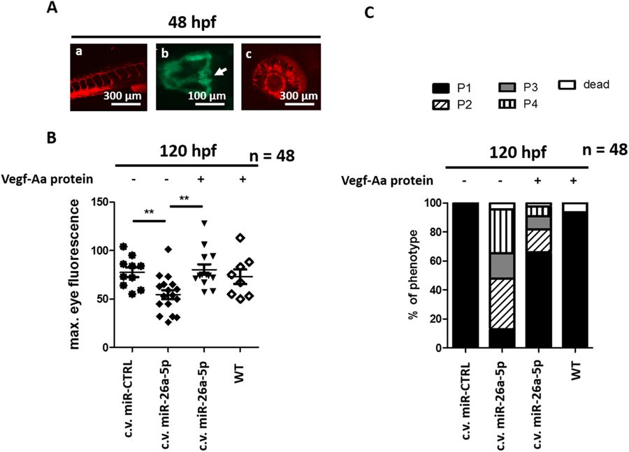
Fig. 5
Vegf-Aa knockdown by miR-26a-5p causes proteinuria and edema in afore healthy zebrafish (A): Pictures of tail vessels (a), glomeruli fusing at zebrafish midline (b) and eye vessel plexus (c) of 48 h old zebrafish shows proper vascular development as well as glomerular fusion. Tail and eye vessel pictures were taken of a Tg(l-fabp:DBP-eGFP/Flk-mcherry) transgenic zebrafish with red fluorescent endothelium. Glomerular fusion is shown in a Tg(wt1b:eGFP) transgenic zebrafish that expresses eGFP under control of the wt1b promotor, arrow points at the fused glomerulus). Scale bar = 100 µM and 300 µM as indicated. (B,C) Quantification of loss of plasma proteins by measuring maximum eye fluorescence of the retinal vessel plexus (B) and quantification of edemous phenotype (C) of Tg(l-fabp:DBP:eGFP) zebrafish larvae at 120 hpf. A control miR mimic (miR-CTRL), a miR-26a-5p mimic (miR-26a-5p) or the combination of miR-26a-5p and Vegf-Aa protein were injected in the cardinal vein at 48 hpf. At this time the tail vasculature, glomerular fusion and retinal vessel formation was already formed; c.v. miR-CTRL n = 10, c.v. miR-26a-5p n = 17, miR-26a-5p + Vegf-Aa n = 13, WT + Vegf-Aa n = 8, total n = 48.