
|
Fig. S3
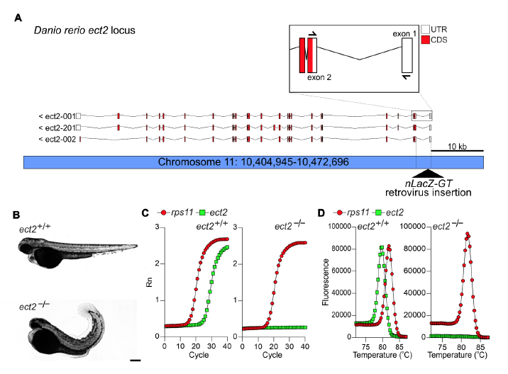
The retroviral insertion hi3820aTg results in disrupted splicing of ect2 mRNA, Related to Figure 2. (A) Schematic representation of the Danio rerio ect2 genomic locus and transcript variants. The hi3820aTg (nLacZ-GT) retroviral integration site is located in the first intron. The retroviral sequence includes splice acceptor sequence and the nLacZ cDNA. Boxed area shows at higher magnification the first three exons and the location of real-time PCR primers used to detect correct splicing of ect2 mRNA. (B) Bright-field images of control (ect2+/+) and ect2hi3820aTg/ hi3820aTg (ect2-/-) embryos at 72 hpf. Mutant embryos exhibit smaller heads and curly tails. (C,D) Real-time PCR amplification (C) and dissociation (D) curves of rps11 (housekeeping control) and ect2 in ect2+/+ and ect2-/- embryos, revealing splicing disruption in ect2 mutant embryos. Scale bar: 200 μm.
Reprinted from Developmental Cell, 44, González-Rosa, J.M., Sharpe, M., Field, D., Soonpaa, M.H., Field, L.J., Burns, C.E., Burns, C.G., Myocardial Polyploidization Creates a Barrier to Heart Regeneration in Zebrafish, 433-446.e7, Copyright (2018) with permission from Elsevier. Full text @ Dev. Cell