
Image
|
Figure Caption
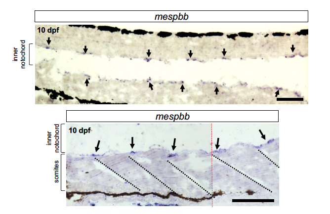
Fig. S3
in situ hybridization of mespbb reveals segmented expression in the notochord sheath, Related to Figure 3.
Cryo-sections of 10 dpf larvae show expression of the mespbb transcript in notochord sheath cells in a segmented pattern (arrows) that correlates with somite boundaries (dotted lines). Scale bars are 50 μm. Red dotted line (bottom panel) indicates where two images were manually stitched together.
Figure Data
Acknowledgments
This image is the copyrighted work of the attributed author or publisher, and
ZFIN has permission only to display this image to its users.
Additional permissions should be obtained from the applicable author or publisher of the image.
Full text @ Cell Rep.