
|
Fig. 4
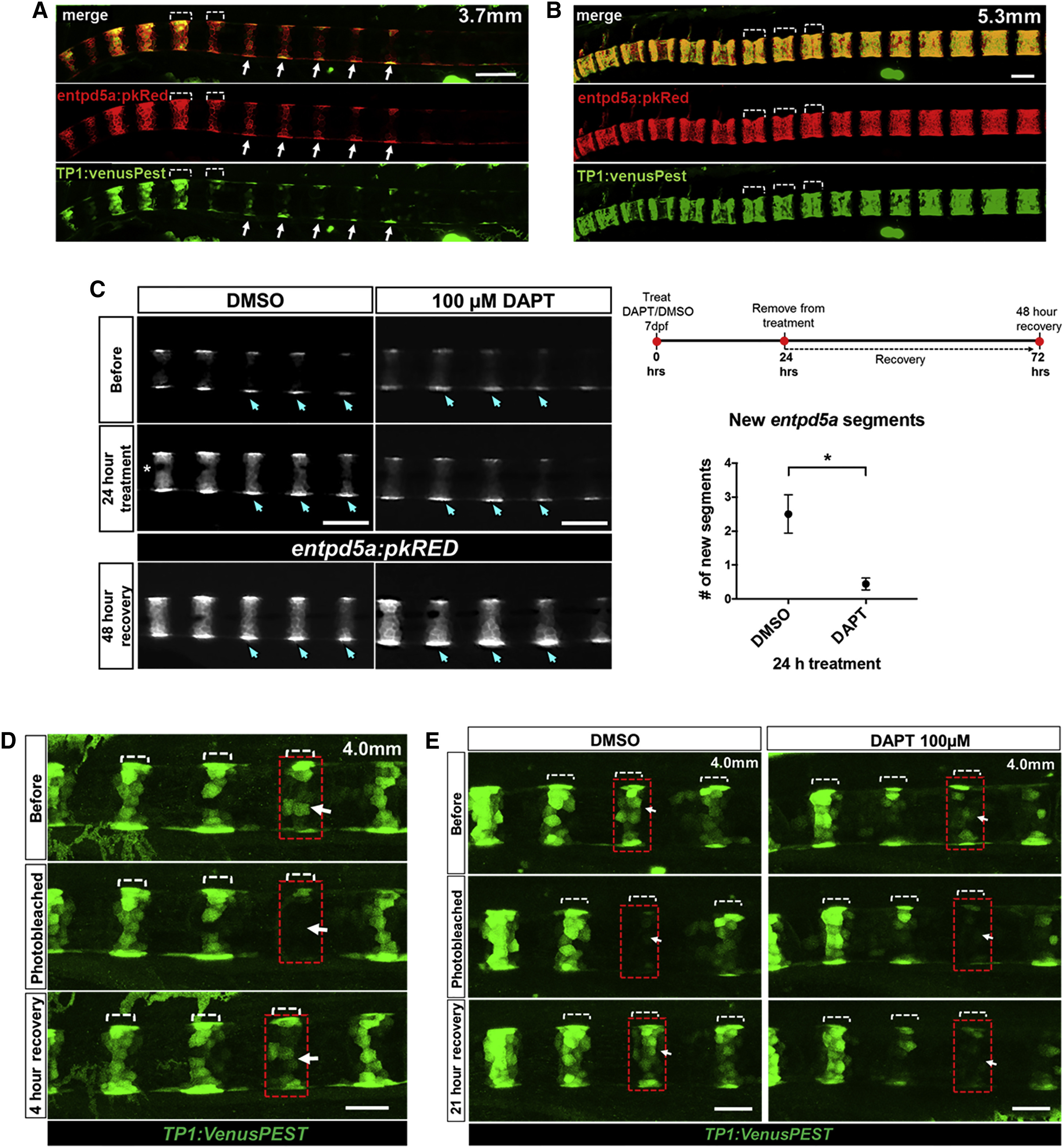
Activation of Notch Signaling in Alternating Sheath Domains Drives Notochord Segmentation
(A) At 3.7 mm SL, expression of entpd5a:pkRED and TP1:VenusPest, a reporter for Notch activation, shows that Notch signaling is active in double-positive and entpd5a+ domains during the formation of new segments (arrows).
(B) At 5.3 mm SL, expression of TP1:VenusPest persists in mature entpd5a+ segments.
(C) Formation of new entpd5a+ segments is halted upon treatment with 100 μM DAPT in 7 dpf larvae (DAPT treated, n = 9; DMSO treated, n = 6; p = 0.013) for 24 hr (see schematic; red dots indicate imaging time point). Control embryos treated with DMSO still form two or three segments (graph). Forty-eight hours following the washout of DAPT, entpd5a+ segment formation is recovered (arrows) in DAPT-treated fish. Scale bars for (A)–(C) are 100 μm.
(D) Photobleaching of a single TP1+ segment (red box) in a 4.0 mm larva shows that TP1 expression is significantly recovered by 4 hr (arrow).
(E) Recovery of the photobleached TP1+ segment (red box) is complete by 21 hr in DMSO but not in 100 μM DAPT. Scale bars for (D) and (E) are 50 μm.