
|
Fig. 1
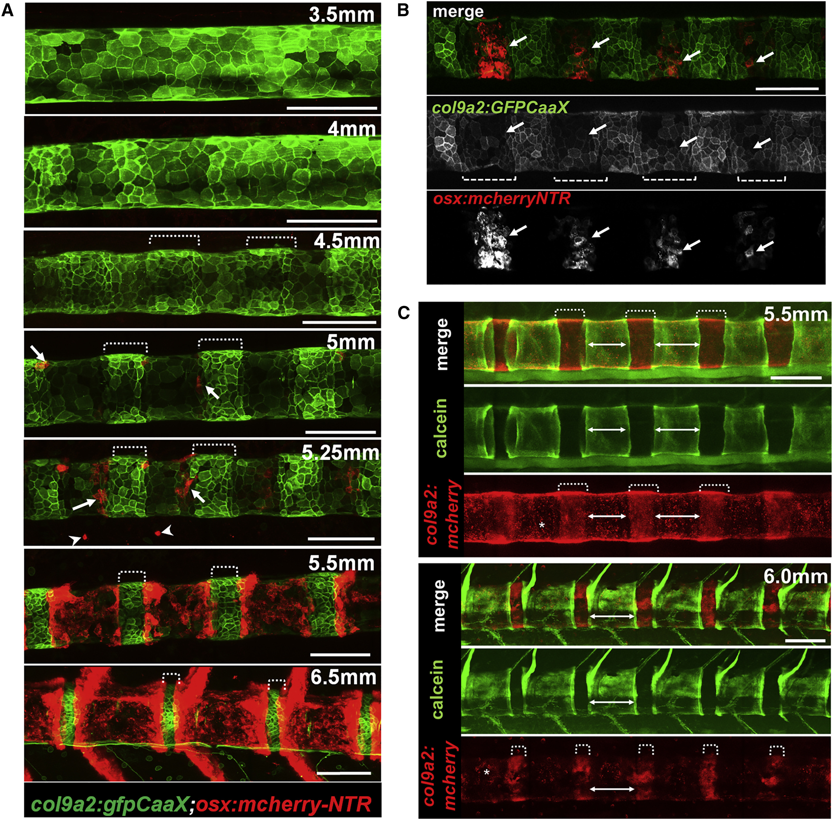
The Notochord Sheath Displays a Segmented Pattern Prior to Vertebral Body Formation
(A) Live confocal imaging of notochord segmentation (denoted by brackets) and osteoblast recruitment (arrows) in col9a2:GFPCaaX and osx:mcherry-NTR fish.
(B) Live confocal imaging showing that osteoblasts specifically migrate to col9a2-negative domains (brackets) in an anteroposterior manner.
(C) Live imaging of calcein stained col9a2:mcherry fish showing that col9a2-negative domains (denoted by asterisks) become mineralized.
Developmental stages are based on standard length. All scale bars are 100 μm. Images in (A) and (C) are digitally stitched.