
|
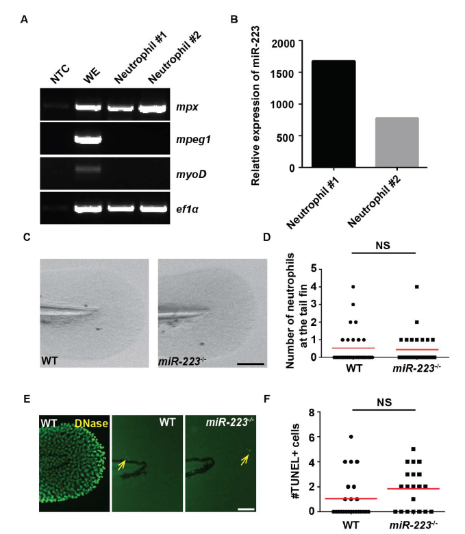
Fig. S1
miR-223 deficiency does not lead to cell death in zebrafish, related to Figure 1.
Neutrophils were isolated by fluorescence-activated cell sorting from 3 dpf zebrafish embryos. (A) RT-PCR of indicated cell markers. mpeg: macrophage marker; mpx: neutrophil marker; myoD: muscle cell marker; ef1a: loading control. WE: whole embryo. NTC: non-template control. (B) RT-qPCR of miR-223 expression in neutrophils, presented as the relative expression to whole embryos normalized to U6. Two individual experiments are shown. (C) Representative images and (D) Quantification of the neutrophil numbers in the fin in WT and miR-223-/- embryos. One representative experiment of three independent repeats is shown. NS, P > 0.05, unpaired student t-test. (E, F) Representative images of TUNEL staining and quantification of TUNEL+ cells in the fin in WT and miR-223-/- embryos. WT embryos treated with DNase I were used as the positive control. Yellow arrows indicate TUNEL+ cells. Scale bars, 100 μm. NS: P > 0.05, unpaired student t-test.