
|
Fig. 10
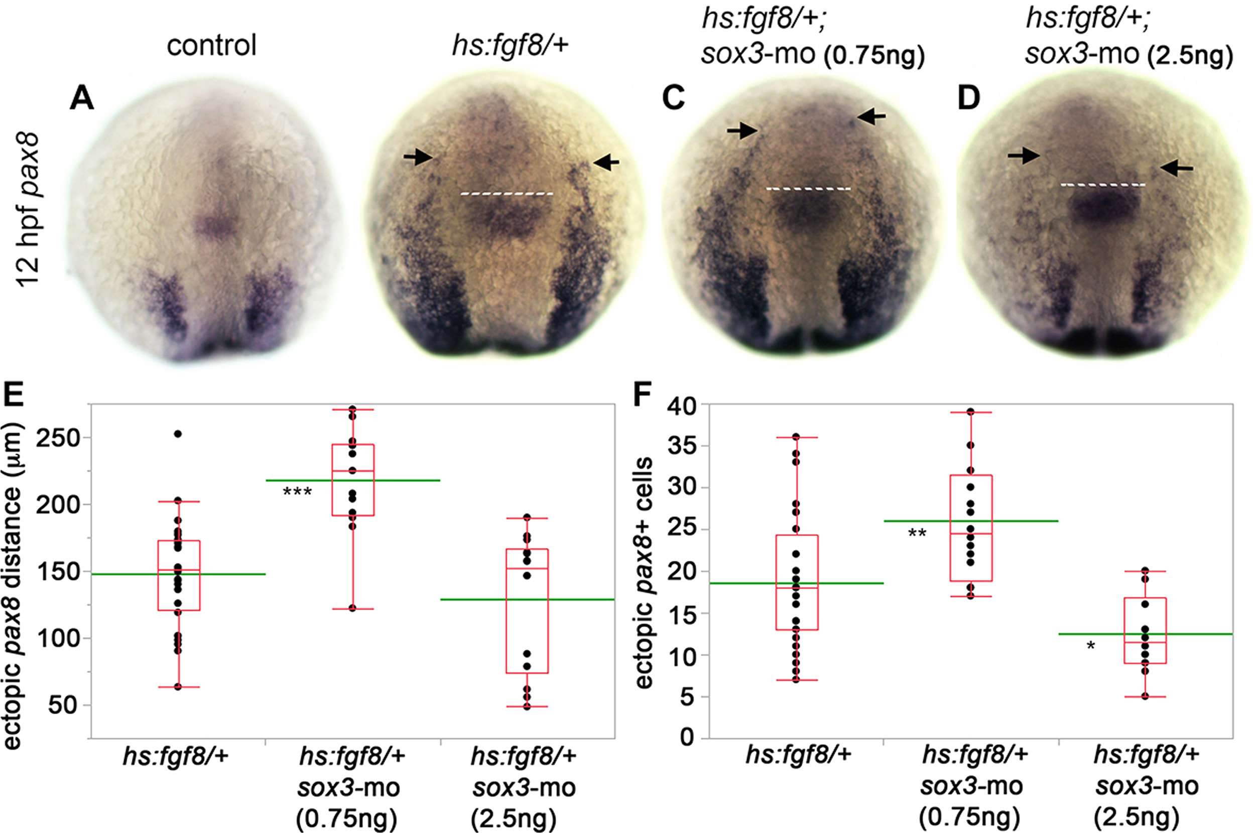
Ectopic otic induction requires an optimal level ofsox3. (A-D) Expression of pax8 at 12 hpf in a control (A), hs:fgf8/+ heterozygote (B), hs:fgf8/+ heterozygote injected with 0.75 ng sox3-mo (C) and hs:fgf8/+ heterozygote injected with 2.5 ng sox3-mo (D) embryo. Embryos were heat shocked at 10 hpf, 39 °C for 30 min. The anterior edge of the midbrain-hindbrain boundary (dashed white line) was used as a reference to measure ectopic expression of pax8. Arrows mark the anterior limit of ectopic pax8 expression (B-D). (E) Box-and-whisker plot of the distance from midbrain-hindbrain border to the anterior limit of ectopic pax8 expression in hs:fgf8/+ heterozygotes, hs:fgf8/+ heterozygotes injected with 0.75 ng sox3-mo and hs:fgf8/+ heterozygotes injected with 2.5 ng sox3-mo. (F) Box-and-whisker plot of the number (per side) of ectopic pax8-expressing cells anterior to the midbrain-hindbrain border in hs:fgf8/+ heterozygotes, hs:fgf8/+ heterozygotes injected with 0.75 ng sox3-mo and hs:fgf8/+ heterozygotes injected with 2.5 ng sox3-mo. The green line indicates the mean. Asterisks indicate statistically significant differences compare to control (* P<0.05, ** P<0.01, *** P<0.001, Tukey's HSD test).
Reprinted from Developmental Biology, 435(1), Gou, Y., Guo, J., Maulding, K., Riley, B.B., sox2 and sox3 cooperate to regulate otic/epibranchial placode induction in zebrafish, 84-95, Copyright (2018) with permission from Elsevier. Full text @ Dev. Biol.