
Fig. 1
- ID
- ZDB-IMAGE-180613-1
- Genes
- Publication
- Lopez-Baez et al., 2018 - Wilms Tumor 1b defines a wound-specific sheath cell subpopulation associated with notochord repair.
- All Figures
- Figures for Lopez-Baez et al., 2018
|
Fig. 1
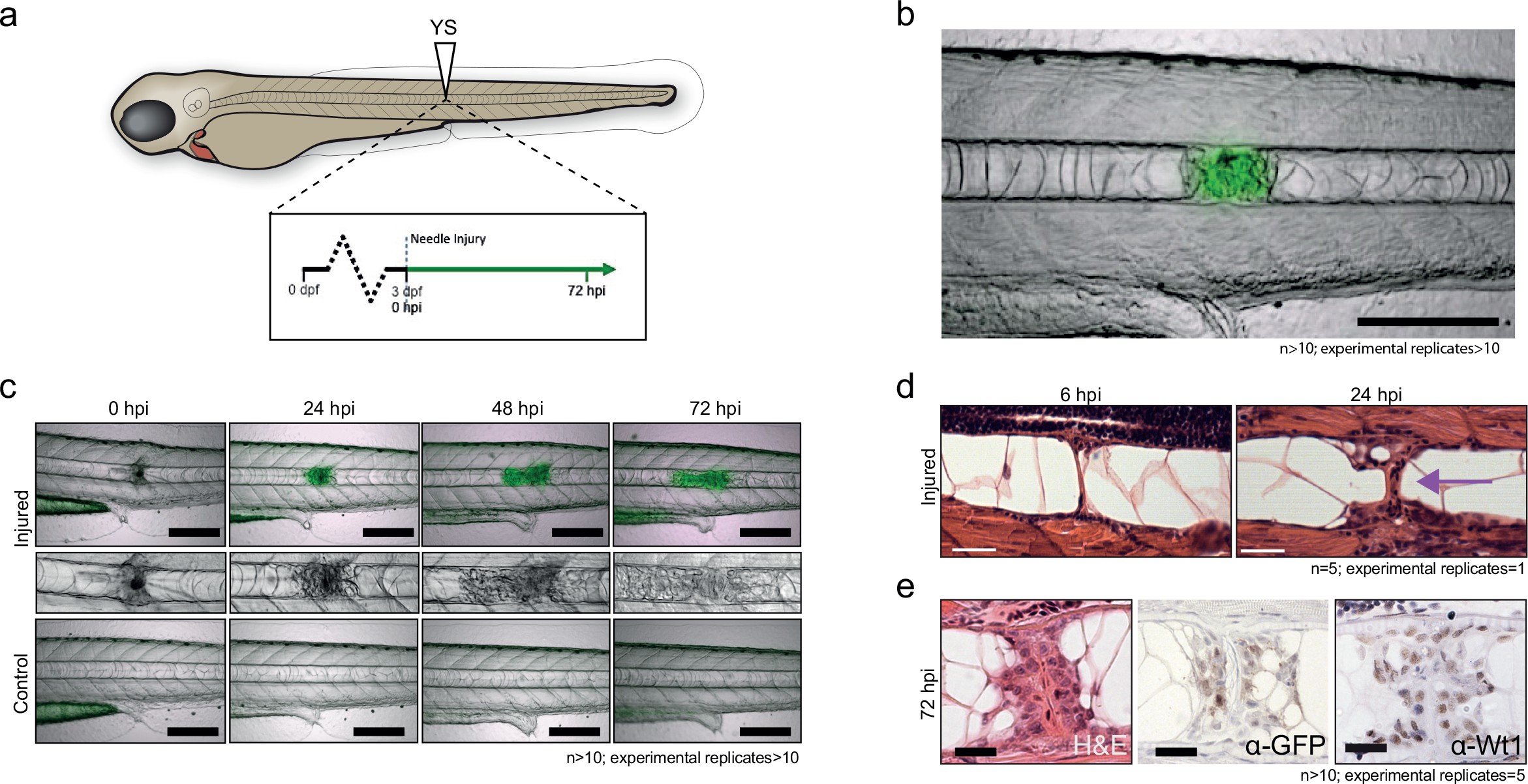
Notochord injury triggers local and sustained wt1b expression.
(a) Schematic of notochord needle-injury protocol. 3 dpf Tg(wt1b:gfp); casper larvae are injured above the yolk sac (YS; at somite 14 or 15) and followed for 72 hr. (b, c) Images of Tg(wt1b:gfp); casper zebrafish trunk over time following notochord needle injury, and uninjured matched controls. GFP signal is associated with a change of cellularity in the injured notochord (inset). n > 10; experimental replicates >10. Scale bar: 100 µm. (d) H and E staining of the injured area at 6 hpi and 24 hpi highlighted the progressive change in cellularity at the site of the injury (arrow). n = 5; experimental replicates = 1. Scale bar: 20 µm. (e) Immunohistochemistry of the injured area with α-GFP and α-Wt1 antibodies. n > 10; experimental replicates = 5. Scale bar: 20 µm. dpf = days post fertilization; hpi = hours post injury; H and E = haematoxylin and eosin.