
|
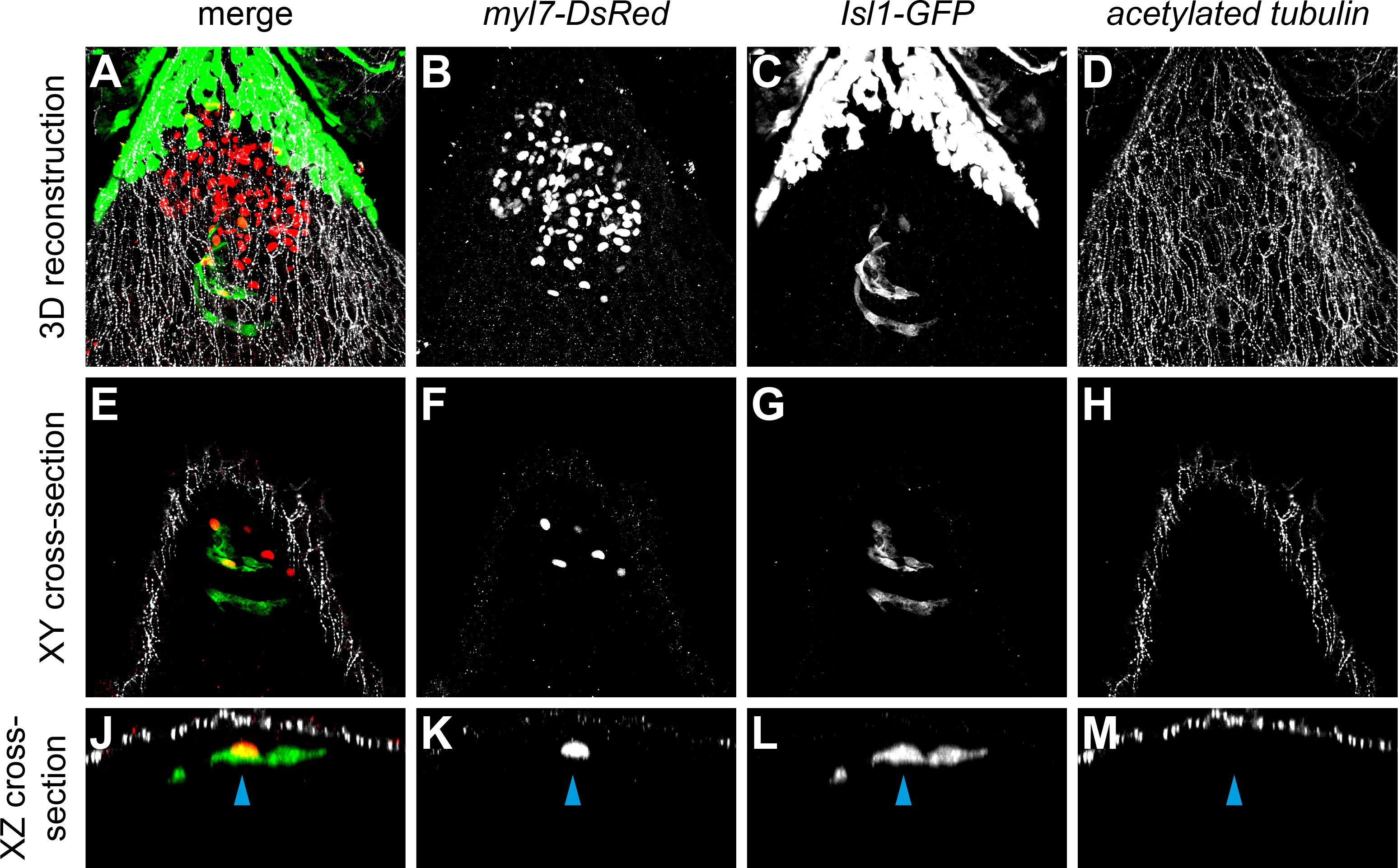
Fig. 6-S2
No neuronal innervation was detected in the 3 dpf heart.
Confocal scans from whole mount embryos containing the tg(myl7-DsRed) (marking all cardiomyocytes) and the tg(Isl1:GFF;UAS:GFP) (reporting isl1 expression and marking pacemaker cells in green) and stained for acetylated tubulin (marking acetylated microtubules found in neurons and axons). Anterior is up and posterior is down. 3D reconstruction (A–D) and cross-sections (E–M). The skin immediately ventral to the heart is covered in axons. The Isl1-GFP-positive pacemaker cells (arrowhead) at the sinoatrial junction are not in direct contact with the acTubulin+ axons.