
|
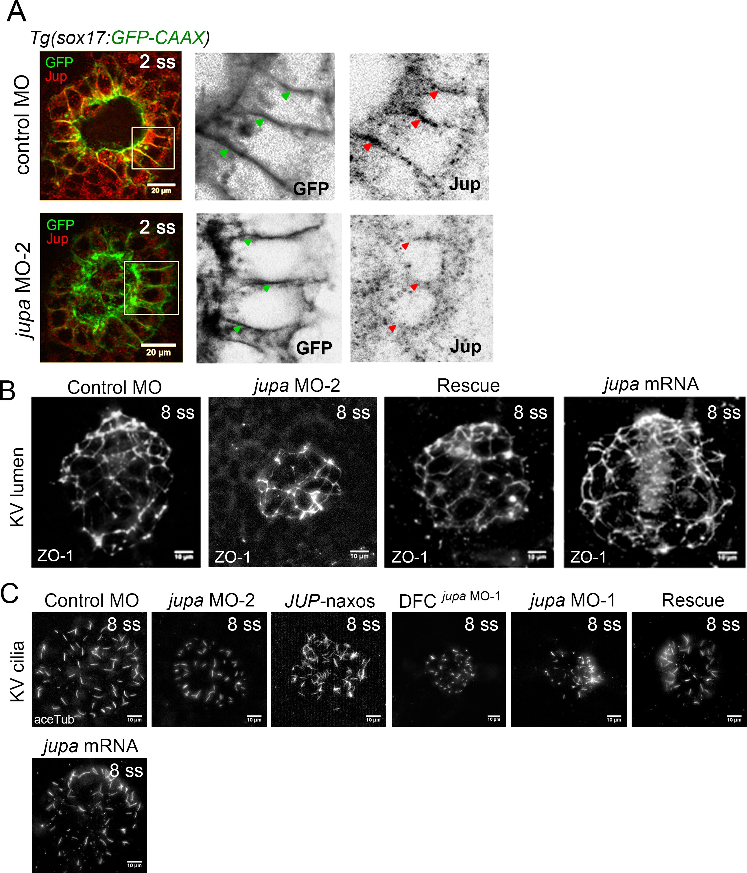
Fig. 5-S2
Interfering with Junction plakoglobin inhibits KV lumenogenesis but not ciliated cell number.
(A) Immunostaining with Jup antibodies showed that embryos injected with jupa MO-2 had reduced Jup protein levels in KV relative to control embryos (image of control MO embryo reproduced from Figure 5A for comparison). Boxes indicate enlarged regions shown as individual channels. Lateral membranes of KV cells are marked by membrane-targeted GFP expression in Tg(sox17:GFP-CAAX) embryos. Arrows point out representative lateral membranes. Scale = 20 μm. (B) Representative KV lumens labeled using ZO-1 antibody staining at 8 ss from different treatments. Scale = 10 μm. (C) KV cilia labeled using acetylated-tubulin antibody staining at 8 ss from different treatments. Scale = 10 μm.