Fig. 6
Fig. 6
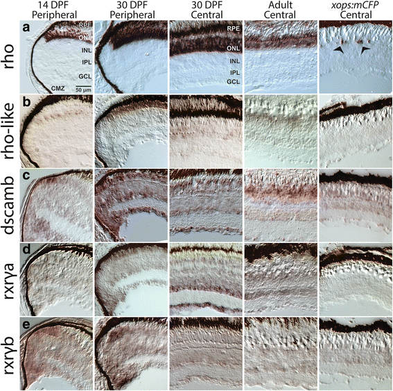
In situ hybridization for transcripts enriched (rho, rhol, dscamb) or present (rxrga, rxrgb) in rods of adult zebrafish, using tissues sampled from larvae (14 days post-fertilization; 14 DPF), juveniles (30 DPF), and adult WT fish, and from xops:mCFP transgenics, which show a chronic rod degeneration [9]. a Expression patterns for rho. Arrows in last panel show degenerating rods in xops:mCFP retina. b Expression patterns for rhol. c Expression patterns for dscamb. d Expression patterns for rxrga. e Expression patterns for rxrgb. DPF, days post-fertilization; CMZ, ciliary marginal zone; RPE, retinal pigmented epithelium; ONL, outer nuclear layer (photoreceptor layer); INL, inner nuclear layer; IPL, inner plexiform layer; GCL, ganglion cell layer. Scale bar (applies to all) = 50 μm

