
|
Fig. 5
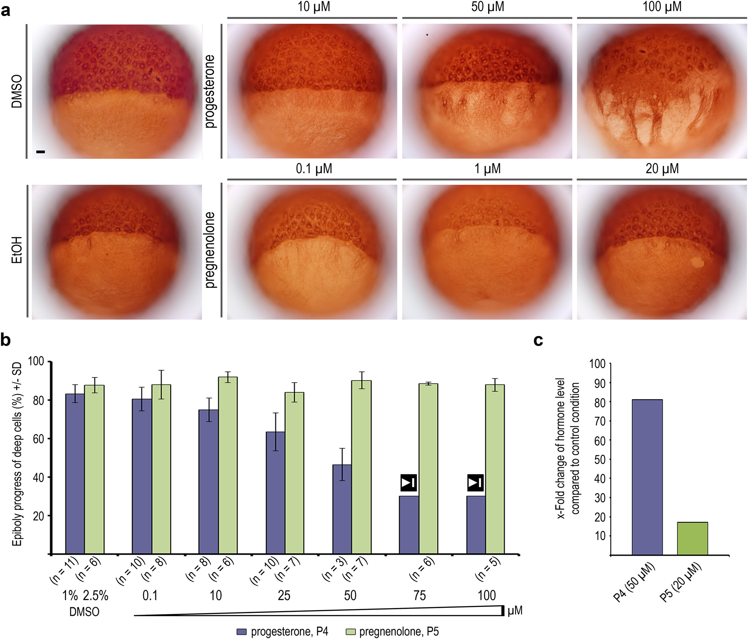
progesterone and pregnenolone effects on YCL microtubules. (a) Anti-β-tubulin immunostaining of wild-type embryos treated with progesterone or pregnenolone at indicated concentrations, and of ethanol or DMSO treated controls. Scale bar 100 µm. (b) Quantification of progesterone and pregnenolone effects on epiboly progression, measured when control embryos reached 80% epiboly (see also supplemental Fig. S3, a and b). Embryos treated with 75 µM or 100 µM progesterone arrested gastrulation at 30–40% epiboly, marked by stop symbol (50 − 100 µM: some pregnenolone precipitation occurred). Error bars - SD. (c) Progesterone and pregnenolone measured by ELISA in wild-type embryos incubated in 50 µM progesterone or 20 µM pregnenolone from 2 hpf onward. Pregnenolone levels increase>10-fold, pregnenolone levels>80-fold.
Reprinted from Developmental Biology, 434(2), Eckerle, S., Ringler, M., Lecaudey, V., Nitschke, R., Driever, W., Progesterone modulates microtubule dynamics and epiboly progression during zebrafish gastrulation, 249-266, Copyright (2017) with permission from Elsevier. Full text @ Dev. Biol.