
|
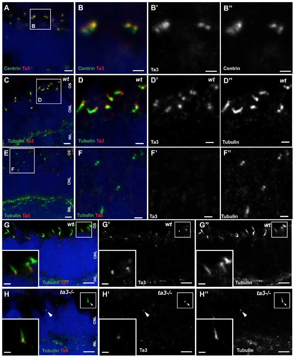
Fig. S1
Ta3 protein localization time course in PRs
(A-B'') 2 dpf retinal cryosections stained with anti-Centrin to mark BBs (green) and anti-Ta3 (red) show localization of Ta3 protein at the BB in wildtype (wt) larvae. (C-D'') 3 dpf cryosections of wt retinas stained with anti-acetylated tubulin and anti-Ta3 3 show localization of Ta3 at both mother and daughter centrioles. (E-F'') In contrast, Ta3 signal is abolished from the majority of BBs in 3dpf ta3 mutants. (G-G'') 4 dpf cryosections of wt retinas stained with anti-acetylated tubulin and anti-Ta3 show similar results as at 3 dpf. (H-H'') In ta3-/- PRs at 4 dpf, the Ta3 signal is mostly abolished (F'') except in a few isolated PRs (arrowhead and arrow in H-H'). Note that the two PRs still expressing Ta3 are also the only ones to have an extended axoneme (arrowhead and arrow in F''). The boxed area in A is shown in B-B'', the one in C is shown in D-D'' and the one in E is shown in F-F''. The insets in G-H'' represent the boxed areas in the corresponding images. Scale bars: 2.5 μm in (A and C), 1 μm in (B-B'', D-D'' and insets in E-F''), 4 μm in (E-F'').