
|
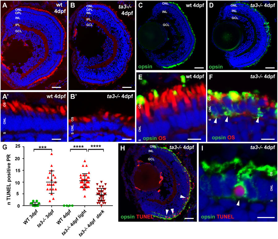
Fig. 2
ta3 mutants show progressive retinal degeneration and intracellular opsin accumulation. (A,B) Retinal lamination is unaffected in ta3 mutants as seen on cryosections at 4 dpf stained with the lipophilic dye BODIPY (red) to mark cell membranes and outer segments and DAPI (blue) to mark nuclei. Note the reduced number of PRs with OSs and the cell shape changes in ta3 mutants compared to wildtype (wt) (A’–B’). (C,D) Marked intracellular opsin accumulation on 4 dpf cryosections stained with 4D2 antibody (green) recognizing rhodopsin and red-green cone opsin on whole eye cryosections. Nuclei are counterstained with DAPI. (E,F) High magnification images of the PR cell layer of wt (E) and ta3 mutant (F) cryosections at 4 dpf stained with 4D2 antibody (green in E,F) and with BODIPY (red in E,F) to highlight the OSs. Note the substantial intracellular opsin accumulation in mutant PRs in (F) (arrowheads). (G) Progressive PR cell death in ta3 mutants as assessed with a TUNEL assay. Note the significantly smaller amount of TUNEL positive cells in 4 dpf ta3−/− larvae raised in darkness (dark red inverted triangles) compared to those raised in a normal light cycle (light red triangles). Quantification was performed on confocal stacks of identical thickness from cryosections of whole retinas through equivalent regions of the eyes. Each datapoint indicates the number of TUNEL positive nuclei counted in a single larva. ***p < 0.001, ****p < 0.0001, Student’s t-test. Bars represent standard deviation. (H) Whole ta3−/− eye cryosection stained with TUNEL assay (red) and with 4D2 antibody (green). Arrowheads mark TUNEL positive PRs. (I) Higher magnification image of a PR with substantial opsin mislocalization (4D2, green) and positive TUNEL reaction (red, arrowhead). dpf days post fertilization, GCL granule cell layer, INL inner nuclear layer, IPL inner plexiform layer, n number, NS not significant, ONL outer nuclear layer, OPL outer plexiform layer, OS outer segment, PR photoreceptor, S synapse, wt wild-type. Scale bars: 30 µm in (A,B) and H, 4 µm in (A’–B’), (E,F) and I, 40 µm in (C,D).