
|
Fig. 1
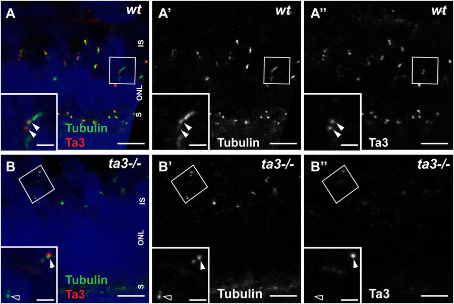
Ta3 localizes to mother and daughter centrioles of photoreceptor primary cilia and is mostly absent in zygotic ta3 mutant PRs. (A–A”) 3 dpf retinal cryosections stained with anti-acetylated tubulin to mark the nascent cilium (green) and with anti-Ta3 (red) show localization of Talpid3 at the base of PR cilia in wildtype (wt) larvae. The double arrowheads highlight Ta3 localization at mother and daughter centriole. (B–B”) In 3 dpf cryosections from ta3−/− larvae, the Ta3 signal is mostly abolished (empty arrowhead in (B”)), but isolated BBs still have positive Ta3 signal, likely maternally deposited (arrowhead in (B”)). The boxed areas in (A–B”) are shown as an inset at the bottom left of each corresponding panel. ONL outer nuclear layer, OS outer segment, S synapse. Scale bars: 2.5 µm in (A–B”), 1 µm in insets.