
|
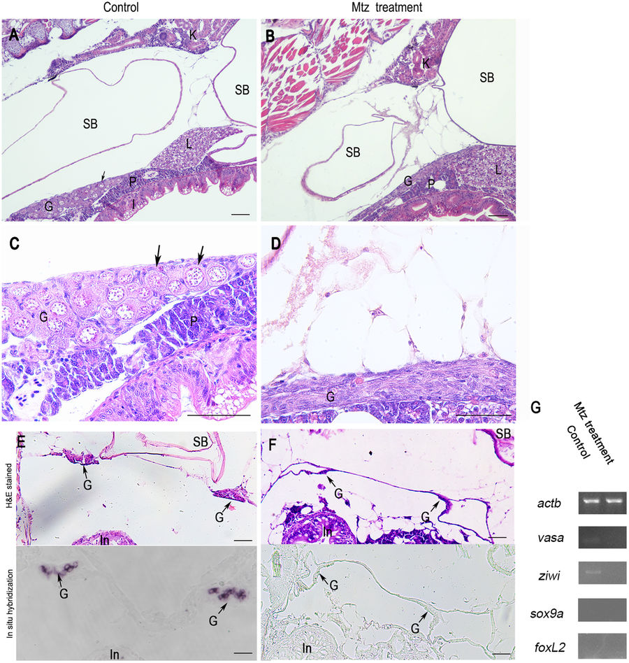
Fig. 3
Germ cells ablation in 20-dpf zebrafish developed from MTZ-treated embryos. Longitudinal sections showing that the gonads of control zebrafish at 20-dpf (A and C) carried a large number of early perinucleolar oocytes (black arrow) with associated somatic tissue, whereas MTZ-treated zebrafish at 20-dpf had only somatic gonadal cells (B and D). Transverse sections showing that the gonads of control zebrafish at 20-dpf (E, upper panel) carried normal gonads (black arrow) with associated somatic tissue and had noraml vasa expression in gonads (E, lower panel, black arrow) detected by in situ hybridization, whereas the gonads of MTZ-treated zebrafish at 20-dpf had only somatic gonadal cells (F, upper panel) and had no vasa expression (F, lower panel) detected by in situ hybridization. (G) RT-PCR results showing the weak expressions of vasa and ziwi were detected in the gonads of control zebrafish but not in the zebrafish developed from the MTZ-treated embryos, and no expressions of sox9a and foxL2 was detetced in neither control zebrafish nor MTZ-treated zebrafish. Expression of actb was used as positive control. K: kidney, SB: swim bladder, G: gonad, L: liver, P: pancreas, I: Intestine. Scale bars = 20 µm (A,B,C,D) and 40 µm (E,F).