
Image
|
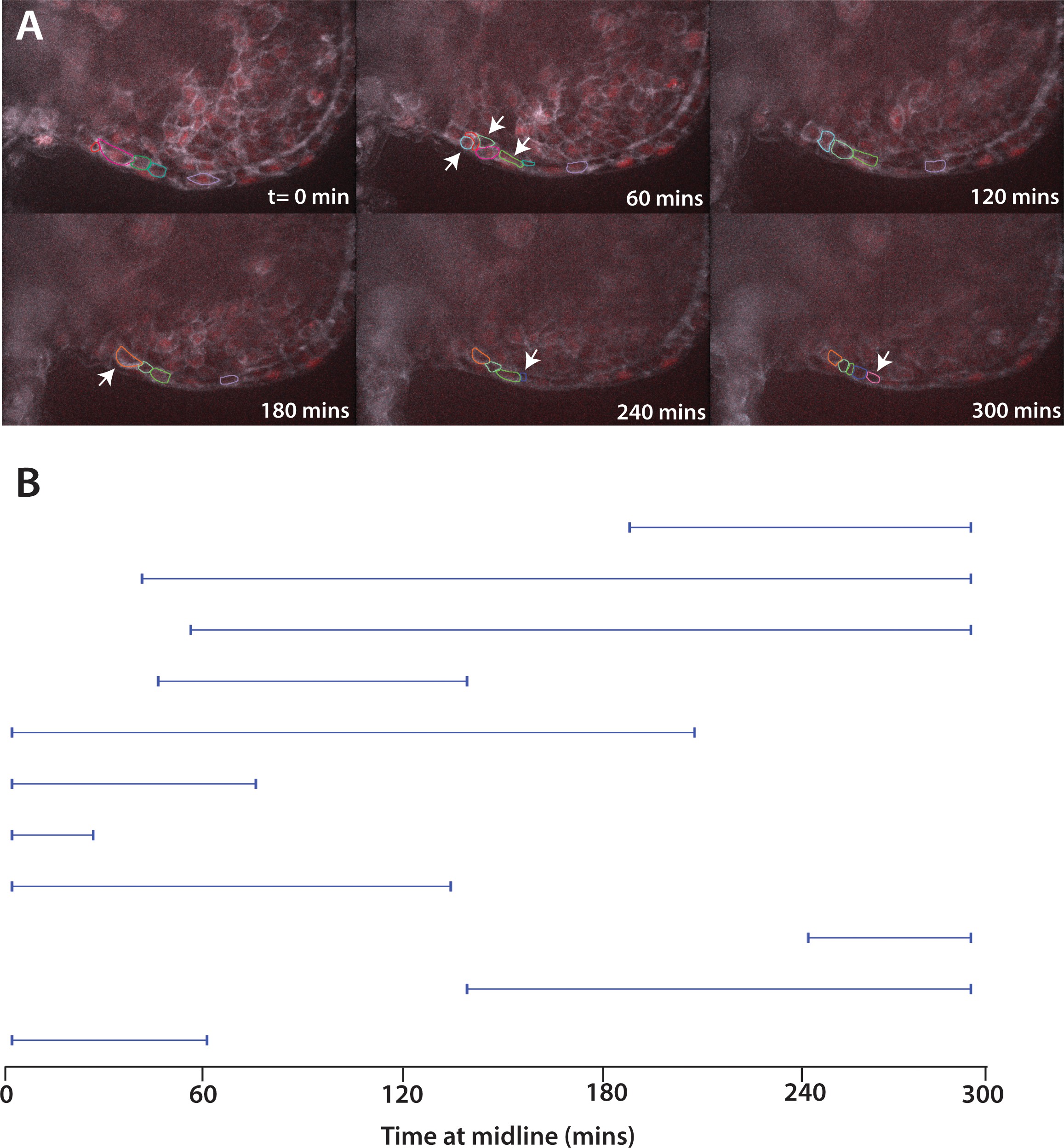
Figure Caption
Fig. 10-S1
Movement of cells expressing Fn∆C.
(A) Stills from live imaging of ventral fin fold formation in embryos expressing Fn∆C from injected mRNA. This movie is a representational movie of one of three movies. Arrowheads show new cells entering the fin fold. (B) The time individual Fn∆C expressing cells were at the midline measured in a single movie. Compare to wild-type cells in Figure 8A. Embryos were positioned on their side and filmed from the left side. When Fn∆C cells crossed over the midline to the right side of the embryo, we stopped recording them, resulting in a truncated line.
Acknowledgments
This image is the copyrighted work of the attributed author or publisher, and
ZFIN has permission only to display this image to its users.
Additional permissions should be obtained from the applicable author or publisher of the image.
Full text @ Elife