
|
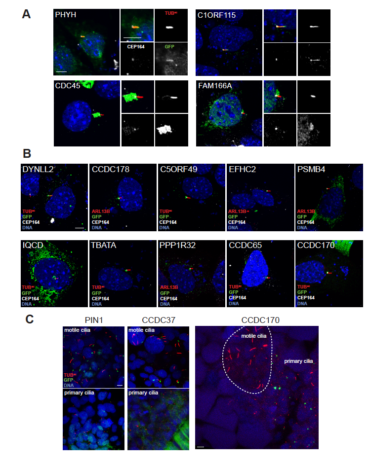
Fig. S1
Subcellular localization of select identified proteins, Related to Figure 3. (A) Immunofluorescence imaging of IMCD3 cell cilia (ARL13B or TUBac, red) and transfected fusions of GFP with randomly selected proteins from the human proteome. The GFP fusions localized to cilia or the ciliary base (green). The distal basal body is marked by CEP164 (white). Nuclei were stained with Hoescht (blue). (B) Immunofluorescence imaging of IMCD3 cells stained as in (A). GFP fusions of the human homologs of randomly selected high-confidence ciliary proteins. (C) Immunofluorescence imaging of three human homologs of randomly selected high-confidence ciliary proteins fused to GFP (green) in the D. rerio embryo (somite stage 6-10). Motile and primary cilia are labeled by TUBac (red) and DNA stained with Hoescht is shown in blue. All scale bars, 5 μm.
Reprinted from Developmental Cell, 43, Sigg, M.A., Menchen, T., Lee, C., Johnson, J., Jungnickel, M.K., Choksi, S.P., Garcia, G., Busengdal, H., Dougherty, G.W., Pennekamp, P., Werner, C., Rentzsch, F., Florman, H.M., Krogan, N., Wallingford, J.B., Omran, H., Reiter, J.F., Evolutionary Proteomics Uncovers Ancient Associations of Cilia with Signaling Pathways, 744-762.e11, Copyright (2017) with permission from Elsevier. Full text @ Dev. Cell