
Image
|
Figure Caption
Fig. S11
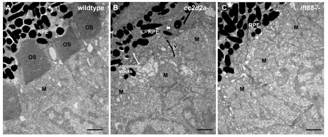
Comparison of cc2d2a and ift88 mutant retinae indicates that vesicle accumulation in PRs is not a general non-specific defect secondary to any ciliary dysfunction.
(A-C) Transmission electron microscopy images of 3 dpf wild-type (A), cc2d2a mutant (B) and ift88 mutant (C) retinae. Note the accumulation of vesicular structures and abnormal membrane stacks in cc2d2a mutants, while no vesicles are found in the inner segments of ift88 mutants.
Acknowledgments
This image is the copyrighted work of the attributed author or publisher, and
ZFIN has permission only to display this image to its users.
Additional permissions should be obtained from the applicable author or publisher of the image.
Full text @ PLoS Genet.