
Fig. S8
- ID
- ZDB-IMAGE-180516-25
- Publication
- Ojeda Naharros et al., 2017 - Loss-of-function of the ciliopathy protein Cc2d2a disorganizes the vesicle fusion machinery at the periciliary membrane and indirectly affects Rab8-trafficking in zebrafish photoreceptors
- All Figures
- Figures for Ojeda Naharros et al., 2017
|
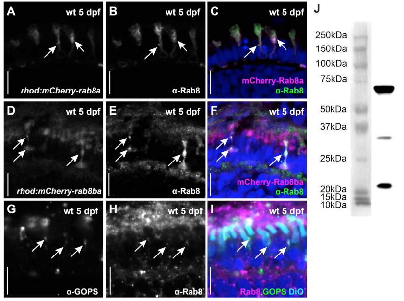
Fig. S8
Comparison of transgenic Rab8 expression with endogenous Rab8 localization.
An anti-Rab8 antibody (B, E and green in C, F) recognizes mCherry-tagged Rab8a (A, magenta in C) and mCherry-tagged Rab8ba (D, magenta in F) on retinal cryosections of 5 dpf zebrafish (arrows). The same antibody (H and magenta in I) recognizes endogenous Rab8 on non-transgenic tissue in a punctated pattern that localizes to the inner segment where it co-localizes with endogenous green opsin (G and green in I, arrows). Anti-Rab8 antibody signal is also very prominent over the retinal pigment epithelium (RPE) but not inside the DiO labelled OSs (cyan in I). Additional puncta are also visible at the synapse and between the nuclei. Such basal punctate localization pattern is partly consistent with mCherry localisation in the transgenic lines which is occasionally seen in such basal regions of the PRs (see time-lapse videos as well). Given that the transgenic lines analyzed express mCherry-Rab8 only in PRs, the relevance of the anti-Rab8 RPE signal cannot be evaluated. (J) A western blot of wild-type whole eyes at 5 dpf probed with the same anti-Rab8 antibody revealed a strong band with the expected size for Rab8 (23.57 KDa). In addition, two other bands were visible on western blot (also acknowledged by the manufacturer: https://www.novusbio.com/products/rab8a-antibody-3g1_h00004218-m02), indicating that the antibody may recognize additional epitopes. Thus, in the absence of Rab8a/Rab8ba/Rab8bb triple knockout mutants, it is not possible to determine the specificity of the anti-Rab8 RPE staining and of the additional puncta with certainty. Scale bars: 10 μm in all panels.