
|
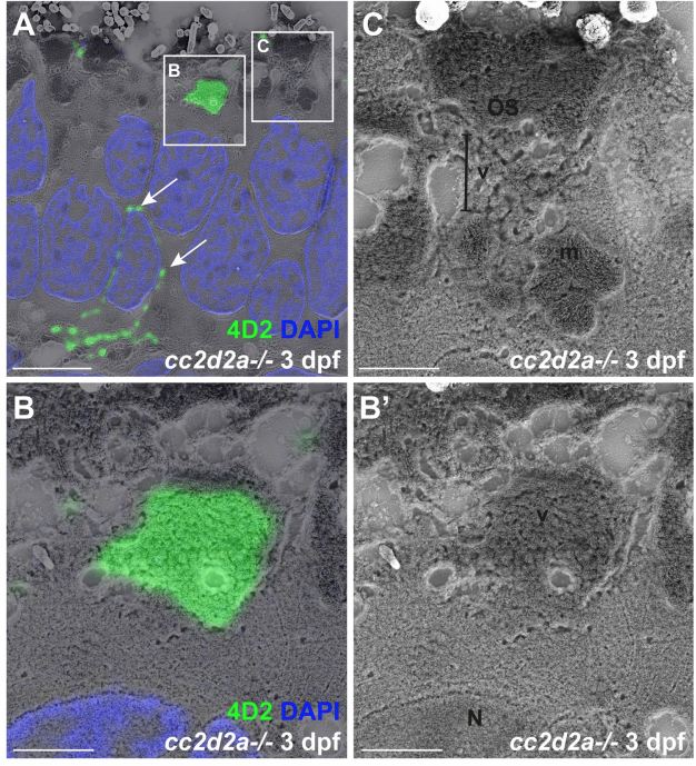
Fig. S3
Accumulated vesicles in cc2d2a-/- PRs at 3 dpf contain opsin.
(A) 3 dpf correlative light and electron microscopy (CLEM) image of a cc2d2a-/- retina stained with 4D2 (green) to label rhodopsin and red-green cone opsin and with DAPI (blue, nuclei). Arrows point to mislocalized opsin inside the cell body. (B-C) Higher magnification images of the boxed regions in (A). (B) Accumulated apical vesicles contain opsin in 4D2-positive PRs. (B’) corresponding scanning electron microscopy image only of (B). (C) Some stacking of membranes in OSs can be occasionally observed above accumulating vesicles. Scale bars: 4 μm in A and 1 μm in B-C. OS outer segment, m mitochondria, N nucleus, v vesicular structures.