
|
Fig. 10
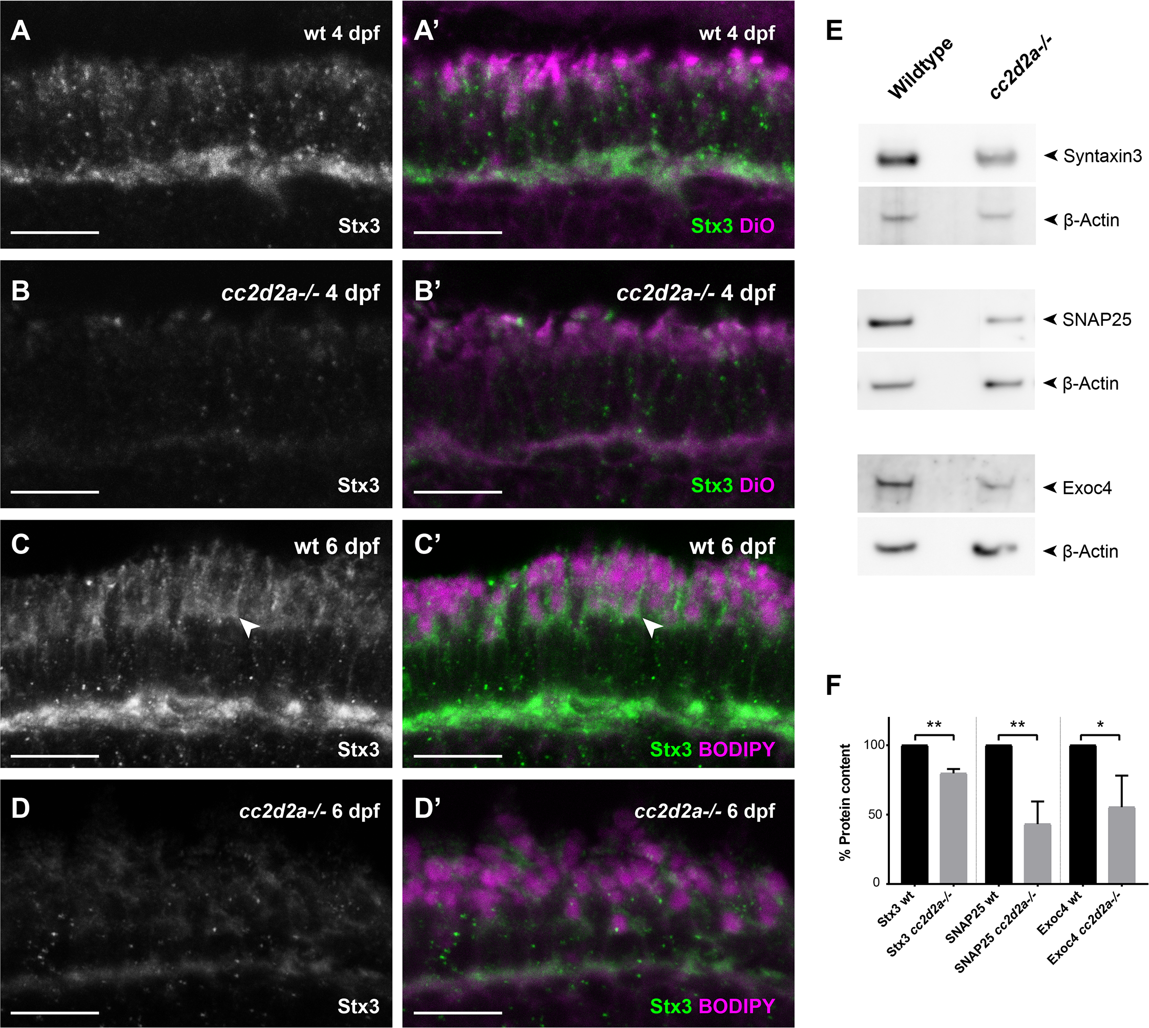
Proteins involved in OCV fusion are affected by loss of Cc2d2a function 4 dpf wild-type (wt) (A-A’) and cc2d2a-/- retinal cryosections (B-B’) and 6 dpf wt (C-C’) and cc2d2a-/- retinal cryosections (D-D’), all stained for Syntaxin3 (Stx3) (grayscale in A-D and green in A’-D’) and counterstained with DiO (A’,B’) or BODIPY (C’,D’) (both magenta) to label membranes. In wt PRs at both developmental times Stx3 localizes along the plasma membrane (arrowhead), between the mitochondrial cluster and the OS and at the synapse (A-A’, C-C’), similar to SNAP25. While minimal Stx3 mislocalization is visible in cc2d2a-/- at 4dpf, a striking decrease in fluorescence intensity is obvious in the mutant (B, D). (E) Western blot on whole eye lysates at 6 dpf confirms decreased protein levels of Stx3 in cc2d2a-/-. Protein levels of SNAP25 and the Exocyst component Exoc4 are also decreased in cc2d2a-/- whole eyes. (F) Relative protein content was determined as the ratio of band intensity relative to the housekeeping protein control (beta-actin), averaged for all replicates and repeated in 3 independent blots. Error bars represent standard deviation. *p<0.05, ** p< 0.01, Student’s t-test. Full western blots are shown in S12 Fig.