
|
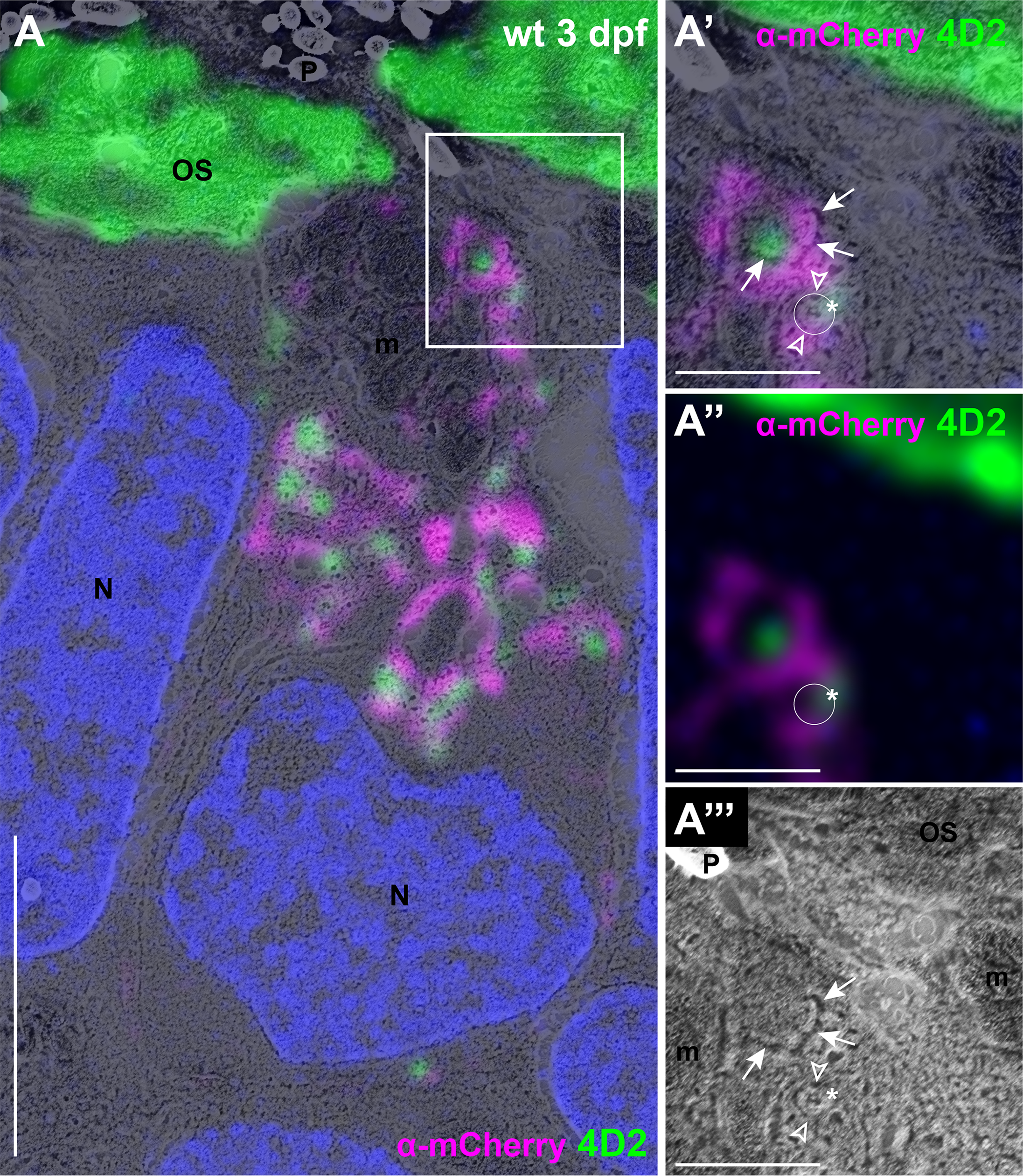
Fig. 4
Rab8 and opsin are associated with membrane-bound vesicular structures (A) CLEM image of wild-type (wt) transgenic zebrafish at 3 dpf expressing mCherry-tagged Rab8a in rods stained with anti-mCherry (magenta) and anti-opsin antibody 4D2 (green).(A’) High magnification image of the boxed area in (A) showing a clearly individualizable small round membrane-bound structure (white circle) coated with Rab8a signal on either side (empty arrowheads over the magenta signal) and with opsin signal over the edge of the structure (asterisk over the 4D2 signal), compatible with transmembrane opsins in a Rab8-coated vesicle. (A”) Immunohistochemistry image only from (A’). The white circle is placed where the vesicular structure is observed in the SEM image. (A”‘) SEM image only from (A’), showing the vesicular structure. The empty arrowheads are placed where the Rab8a signal is observed and the asterisk is located over the opsin signal. The additional m-Cherry and opsin signal likely represent a conglomerate of several vesicular structures with multilobulated membranes (arrows in A’ and A”‘). Scale bars: 4 μm in A and 1 μm in A’-A”‘. m mitochondria, N nuclei, OS outer segments, P pigment, wt wild-type.